
Review began 08/28/2023 Review ended 09/11/2023 Published 09/16/2023
Open Access Original
Article DOI: 10.7759/cureus.45386
The Role of Computed Tomographic Angiography in Predicting the Development of Vasospasm Following Ruptured Intracranial Aneurysm Microsurgery
© Copyright 2023
Varol. This is an open access article distributed under the terms of the Creative
Eyüp Varol
1
Commons Attribution License CC-BY 4.0., which permits unrestricted use, distribution, and reproduction in any medium, provided the original author and source are credited.
1. Neurological Surgery, Umraniye Training and Research Hospital, Istanbul, TUR
Corresponding author: Eyüp Varol, dreyupvarol@gmail.com
Abstract
Introduction
Following subarachnoid hemorrhage, cerebral vasospasm is the primary cause of morbidity and death. The aim of this study is to predict the development of vasospasm by detecting changes in vessel diameter after surgery using computed tomography angiography.
Methods
We retrospectively evaluated the patients who underwent aneurysm clipping due to a bleeding aneurysm between 2019-2022. Age, gender, location, subarachnoid hemorrhage grades, development of perioperative rupture, and temporary clip use were examined. Preoperative and postoperative diameters of the internal carotid artery, A1-A2, and M1-M2 were measured. Radiological and clinical vasospasm development in the postoperative period was also documented.
Results
The aneurysm localizations of the 100 patients (mean age: 50.38±13.04 years) were anterior cerebral artery in 50 patients, internal carotid artery in 37 patients, and middle cerebral artery in 30 patients. In the postoperative follow-up, radiological vasospasm was apparent in 41 patients. The changes in arterial diameter reveal a statistically significant decrease in the internal carotid artery, M1-M2, and A1-A2 artery diameters on the operated side compared to the contralateral side (p<0.001). Based on the receiver operating characteristic (ROC) analysis, the most likely change in arterial diameter on the operated side to indicate the presence of vasospasm was calculated from the available data, where the decrease in total arterial diameter was 13.7%.
Conclusion
Vasospasm remains one of the significant causes of morbidity and mortality post subarachnoid hemorrhage. While there have been advances in imaging modalities, predicting which patients will develop vasospasm has remained elusive. Our research attempts to provide a quantifiable metric (13.7% decrease in vessel diameter) that can be an early predictor of this complication.
Categories: Neurosurgery
Keywords: computed tomographic angiography, vasospasm, hemorrhage, subarachnoid, aneurysm
Introduction
Aneurysmal subarachnoid hemorrhage (aSAH) is a life-threatening disease that affects three to 25 people per 100,000 worldwide every year [1]. Vasospasm is evident in around 40% of aSAH patients, and 20-30% of aSAH patients suffer from vasospasm-related neurological impairments [2-5]. Following subarachnoid hemorrhage (SAH), cerebral vasospasm (CVS) is one of the common causes of morbidity and death [6]. Vasospasm affects 50-70% of SAH patients, with 50% of these patients experiencing neurological symptoms (i.e., symptomatic CVS) [7]. A new focal neurological deficit that is not explained by rebleeding or hydrocephalus and an altered level of consciousness are common indications of vasospasm. CVS is well known to cause neurological impairments through delayed cerebral ischemia [3,8,9].
Cerebral infarction affects half of all symptomatic CVS patients and is deadly in 30% of cases [10]. It is critical to research CVS to support the development of effective therapies and reduce the morbidity rate of people with this ailment. Despite efforts to develop novel medicines to prevent and cure CVS, it continues to be a major cause of death and mortality in patients who survive initial aSAH therapy [11,12]. One of the objectives of critical care monitoring in these patients is early diagnosis of CVS. Cerebral digital subtraction angiography (DSA) is currently the gold standard for diagnosing CVS [13,14]. Nevertheless, it is not apparent
How to cite this article
Varol E (September 16, 2023) The Role of Computed Tomographic Angiography in Predicting the Development of Vasospasm Following Ruptured Intracranial Aneurysm Microsurgery. Cureus 15(9): e45386. DOI 10.7759/cureus.45386
if these data might be comparable to the DSA resolution images, which are the gold standard imaging
technique [15]. Magnification, the distance from the source of the image to the detector, and the viewing
angle are some of the variables that influence vessel diameter measurements on DSA. Furthermore,
measurements are frequently provided only in relative units when assessing pictures with simple imaging
viewers [16].
Transcranial Doppler (TCD) ultrasonography is one example of the non-invasive methods that can be used
to detect vasospasm in patients with aSAH. However, its low specificity and high operator dependence
render it an inadequate detection tool for vasospasm [17]. Another non-invasive imaging technique,
computed tomographic angiography (CTA), shows greater promise as a more accurate tool for evaluating
vasospasm. This established routine screening tool provides non-invasive information about cerebral artery
diameters [18]. Multidetector CTA, which enables rapid image acquisition and lower radiation exposure, has
gained popularity over the last decade. Our aim in this study is to use CTA to evaluate changes in vessel
diameter after surgery and assess the effects of those changes on vasospasm in order to predict the
development of vasospasm.
Materials And Methods
Patient data and outcome assessment
For this research, we retrospectively evaluated the collected data of patients in our clinic who underwent
aneurysm clipping due to a bleeding aneurysm between 2019 and 2022.
Inclusion criteria
The inclusion criteria included patients who underwent aneurysm clipping due to a bleeding aneurysm
between 2019 and 2022; patient age greater than 18 years and less than 85 years; availability of both
preoperative and immediate postoperative CTA and diffusion-weighted images (DWI) data; patients with
complete clinical documentation, including recorded data about age, gender, aneurysm location, Fisher
classification, Hunt-Hess (HH) classification, and World Federation of Neurosurgical Societies (WFNS)
grade; and patients who provided written informed consent.
Exclusion criteria
The exclusion criteria included patients with incomplete clinical documentation or missing preoperative or
postoperative imaging data; patients with other cerebral pathologies such as tumors, arteriovenous
malformations, or previous strokes which could interfere with accurate measurement of arterial diameters;
patients who did not provide informed consent or those unable to provide consent due to incapacitation;
patients with other surgical interventions or endovascular procedures on the cerebral arteries within the
previous year; patients with contraindications to CTA or DWI such as severe allergy to contrast agents or
metallic implants; pregnant women or those nursing; and patients with chronic kidney disease or renal
dysfunction, which contraindicates the use of contrast agents.
The recorded data concerns the patient’s age and gender, the location of the aneurysm causing subarachnoid
bleeding, Fisher classification, Hunt-Hess (HH) classification, WFNS grade, single or multiple aneurysm,
development of perioperative rupture, and temporary clip use and duration. The recorded blood volumes in
the aspirator were used to determine the amount of bleeding during the operation. Preoperative and
postoperative vessel diameters of the internal carotid artery (ICA), A1-A2, and M1-M2 were measured on
the operated and non-operated sides and noted. All measurements were made by two experienced
neurosurgeons, and their mean values were taken. The development of radiological and clinical vasospasm
in the postoperative period was also documented. If a newly developed restriction was observed on DWI, it
was evaluated as a radiological vasospasm. It was accepted as a clinical vasospasm if a newly onset
neurological deficit was observed. Patient data are reported according to common descriptive statistics.
Written informed consent was obtained from all patients, and the study was performed in accordance with
the ethical standards of the Declaration of Helsinki and under the approval of our institutional review
committee. Ethical approval for the study was obtained from the Umraniye Training and Research Hospital
Ethics Committee.
Radiological technique
The same tomography device was employed for all patients in the study. Preoperative and postoperative
vessel diameters were measured in all patients by two senior neurosurgeons, and the assessment used the
average of the measurements taken by the two neurosurgeons. Preoperative and immediate (<24 hours)
postoperative CTA and DWI were conducted according to a standard predefined imaging protocol. The
vascular diameters were assessed in preoperative and postoperative CTA images on operated and non
operated parts. Measurements were performed at 1 cm distal to the anterior cerebral artery (ACA) origin, 1
cm distal to the M1 and M2 origins, and 1 cm proximal to the formation of the ACA and middle cerebral
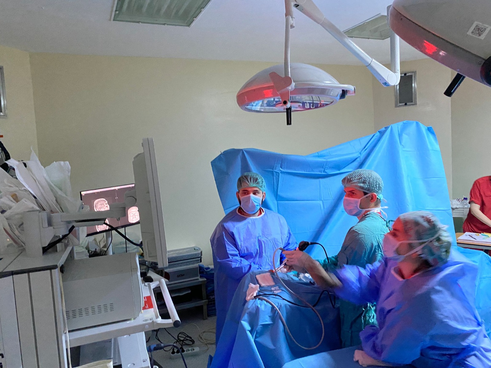
artery (MCA) composition for the ICA (Figure 1).
2023 Varol et al. Cureus 15(9): e45386. DOI 10.7759/cureus.45386 2 of 9


FIGURE 1: Measurements of arterial diameters in a 3D image viewer
software
a) internal carotid artery, b) M1 segment of middle cerebral artery, c) A1 segment of anterior cerebral artery, d) A2
segment of anterior cerebral artery
Preoperative and postoperative DWI were performed in all patients for radiological evaluation of vasospasm.
Statistical analysis
The Statistical Package for the Social Sciences (SPSS) version 25.0 (IBM Inc., Armonk, New York) program
was used for statistical analysis to evaluate the results of this study. Descriptive, graphical, and statistical
methods were applied to determine whether the scores obtained from each continuous variable were
normally distributed. The Kolmogorov-Smirnov test was used to test the normality of the scores obtained
from a continuous variable with the statistical method. Descriptive statistical methods (number, percentage,
mean, median, standard deviation, etc.) were used while evaluating the research data. Comparisons between
two groups in quantitative data were made with the independent samples t-test (in data with normal
distribution) and Mann-Whitney U test (in data with no normal distribution), and comparisons of more than
two groups were made with the Kruskal-Wallis test. The Bonferroni test was used to determine from which
groups the difference originated, while Chi-squared tests (Pearson’s chi-squared test, continuity correction
test, and Fisher’s exact test) were applied for qualitative comparisons between groups. Repeated
measurements were made with the paired samples t-test. Receiver operating characteristic (ROC) analysis
was used to determine the most appropriate rate of change in arterial diameter in the presence of
vasospasm. A p-value of less than 0.05 was considered statistically significant with a 95% confidence
interval.
Results
Within the scope of the study, the findings of 100 patients were analyzed. The mean age of the patients was
50.38 ± 13.04 years, and 50% of them were in the age group of 50 years and above. Of the 100 patients, 51%
were female, and 49% were male. According to radiological imaging results, the aneurysm localization was
ACA in 50 patients, ICA in 37 patients, and MCA in 30 patients. Aneurysm was observed as multiple in 16
patients and single in 84 patients. Temporary clips were applied to 47% of the patients. The presence of
perioperative rupture was reported in 34% of the patients, and the mean amount of perioperative bleeding
was 306.20 ± 191.92 mL. In the postoperative follow-up, radiological vasospasm was apparent in 41 patients.
During the treatment period, morbidity was observed in 15 (15%) patients, and mortality was observed in 13
(13%) patients (Table 1).
2023 Varol et al. Cureus 15(9): e45386. DOI 10.7759/cureus.45386 3 of 9

| n=% Mean(SD) Min.-Max. Age Total 100 50.38 (13.04) 24-79 <50 50 Age group ≥50 50 Male 49 Gender Female 51 Right 55 Surgery side Left 45 ACA 50 Aneurysm location ICA 37 MCA 30 Single 84 Aneurysm single/ multiple Multiple 16 Yes 47 Temporary clip No 53 Yes 34 Perioperative rupture No 66 Yes 41 Vasospasm No 59 Perioperative bleeding (cc) Total 100 306.20 (191.92) 50-1000 Discharged with healing 72 Discharge situation Need care 15 Dead 13 |
TABLE 1: Patient demographics (N=100)
ACA – anterior cerebral artery, ICA – internal carotid artery, MCA – middle cerebral artery, Min – minimum, Max – maximum, SD – standard deviation
We examined the WFNS, Fisher, and HH scale results of the patients and found that the mean scores were
2.02 ± 1.30, 2.59 ± 1.19, and 2.12 ± 1.17, respectively.
There was no statistically significant difference between the measurements made by the two neurosurgeons.
The changes in arterial diameter reveal a statistically significant decrease in the ICA, M1-M2, and A1-A2
artery diameters on the operated side compared to the contralateral side (p<0.001). The evaluation of all
arteries together found a 13.2% decrease in mean arterial diameter on the operated side and a 3% decrease
on the opposite side (p<0.001). In the preoperative period, the total arterial diameter was higher on the
operated side (2.40 ± 0.27) than on the contralateral side (2.29 ± 0.23) to a statistically significant extent
(p=0.004). In the postoperative period, a decrease in arterial diameter was detected on the operated side
(2.08 ± 0.30) compared to the contralateral side (2.22 ± 0.20), also with statistical significance (p<0.001;
Table 2).
2023 Varol et al. Cureus 15(9): e45386. DOI 10.7759/cureus.45386 4 of 9

| Preoperative Postoperative Difference %↓ Artery diameter (mm) Side Mean±SD Mean±SD p-value Mean±SD Operated 3.34±0.49 2.92±0.53 <0.001a* 12.5±8.9 ICA Non-operated 3.14±0.42 3.08±0.40 <0.001a* 1.7±4.2 p-value 0.002c* 0.018c* <0.001b* Operated 2.07±0.28 1.80±0.31 <0.001a* 12.7±9.8 Non-operated 2.03±0.24 2.01±0.22 0.009a* 1.0±4.2 M1-2 p-value 0.332c <0.001c* <0.001b* Operated 1.78±0.22 1.51±0.23 <0.001a* 14.7±12.0 A1-2 Non-operated 1.68±0.21 1.54±0.20 <0.001a* 7.7±10.0 p-value <0.001c* 0.397c <0.001b* Operated 2.40±0.27 2.08±0.30 <0.001a* 13.2±8.3 Total Non-operated 2.29±0.23 2.22±0.20 <0.001a* 3.0±4.3 p-value 0.004c* <0.001c* <0.001b* |
TABLE 2: Change in artery diameters before and after surgery (N=100)
* p<0.05, a – paired samples t-test, A1-2 – A1-2 segments of anterior cerebral artery, b – Mann-Whitney U test, c – independent samples t-test, ICA – internal carotid artery, M1-2 – M1-2 segments of middle cerebral artery, SD – standard deviation
While not statistically significant (p=0.364), the rate of vasospasm was higher in patients with perioperative
temporary clips than in those without (47% vs. 36%). The rate of vasospasm was also higher in patients with
perioperative rupture than in those without (56% vs. 33%), which was at the limit of statistical significance
(p=0.050). The rate of vasospasm was 32% in discharged patients, 80% in patients who needed care, and 46%
in patients who died (p=0.002).
The rate of vasospasm was found to be higher among those with a Fisher grade of III or above than among
those with a grade of II or below (51.7% vs. 25%, p=0.014). The rate was also higher in patients with a WFNS
score and HH classification of II or above compared to those with a score below II (WFNS: ≥II, 59.6% vs. <II,
20.8%, p<0.001; H&H: ≥II, 50% vs. <II, 21.9%, p=0.014). The Fisher grade, WFNS, and HH classification
scores of patients with vasospasm were higher than those of patients without vasospasm. This difference
was statistically significant (p<0.05). Among patients presenting with vasospasm, there was no statistically
significant difference in terms of age or amount of perioperative bleeding (p>0.05; see Table 3).
2023 Varol et al. Cureus 15(9): e45386. DOI 10.7759/cureus.45386 5 of 9

| Vasospasm p-value Yes (n=41), mean±SD No (n=59), mean±SD Age 50.71±13.69 50.15±12.68 0.763 Bleeding (cc) 332.20±214.65 288.14±174.02 0.325 Fisher grade 3.00±1.00 2.31±1.24 0.005c* WFNS grade 2.32±1.21 1.81±1.33 0.002c* Hunt-Hess grade 2.34±1.11 1.97±1.20 0.020c* |
TABLE 3: Averages of some continuous variables according to the presence of vasospasm (N=100)
* p<0.05 with Mann-Whitney U test, SD – standard deviation, WFNS – World Federation of Neurosurgical Societies
Compared to patients without vasospasm, patients with vasospasm showed a greater, statistically significant
decrease in arterial diameter on both the operated and contralateral sides, ICA, M1-M2, A1-A2, and total
arterial diameters (p<0.05). In patients with perioperative rupture, there was a greater decrease in arterial
diameter on both the operated and contralateral sides, ICA, A1-A2, and total arterial diameters compared to
patients without perioperative rupture. This difference was statistically significant (p<0.05).
The area under the curve was found to be 0.967 (95% CI: 0.931-1); accordingly, the change in arterial
diameter (% decrease rate) was found to be statistically significant (p<0.001) for determining the presence of
vasospasm. Based on the ROC analysis, the most likely change in arterial diameter on the operated side to
indicate the presence of vasospasm was calculated from the available data, where the decrease in total
arterial diameter was 13.7%. For the cutoff value of 13.7%, the sensitivity was 100%, the specificity was
91.5%, the positive predictive value was 89.1%, the negative predictive value was 100%, and the overall
accuracy was 95% (Table 4).
| Radiological vasospasm occurrence Cutoff value 13.7% AUC (%95 CI) 0.967 (0.931-1) p-value <0.001 Sensitivity 100% (41/41) Specificity 91.5% (54/59) PPV 89.1% (41/46) NPV 100% (54/54) Accuracy 95% (95/100) |
TABLE 4: Optimal positive cutoff limit for the rate of decrease in total artery diameter of the operated side in determining the presence of vasospasm (ROC analysis results)
AUC – the area under the ROC curve, CI – confidence interval, NPV – negative predictive value, PPV – positive predictive value
Discussion
Our study aimed to bridge a critical gap in the field of neurosurgery by investigating the utility of CTA in
predicting the development of vasospasm in patients with aSAH after surgery. The primary outcomes of our
study reveal a statistically significant decrease in arterial diameters, particularly in the ICA, M1-M2, and A1-
A2 arteries on the operated side compared to the contralateral side. These findings suggest that CTA can be a
valuable tool for monitoring postoperative changes in vessel diameter, potentially identifying patients at a
2023 Varol et al. Cureus 15(9): e45386. DOI 10.7759/cureus.45386 6 of 9

higher risk of developing vasospasm. This information can enable clinicians to take proactive measures,
such as closer neurological monitoring and targeted interventions, to prevent or mitigate the impact of
vasospasm.
Moreover, the ability to predict vasospasm early in the postoperative period can significantly improve
patient outcomes. It can lead to timely interventions that reduce the risk of cerebral infarction and its
associated morbidity and mortality. Our study lays the foundation for further research in this area. It may
contribute to developing standardized protocols for using CTA in the postoperative monitoring of aSAH
patients. The ability to predict vasospasm early and take proactive measures based on CTA measurements
can improve patient care and reduce the devastating consequences of vasospasm following aSAH.
The main cause of focal cerebral ischemia after SAH is CVS [19]. While rebleeding is the most frightening
complication that can develop after aSAH, it has gradually decreased in prominence due to the widespread
practice of early surgery. Meanwhile, vasospasm has become the most risky complication of SAH in terms of
mortality and morbidity [3,8,9]. Therefore, early recognition of vasospasm is vital. Aneurysmal SAH patients
with a history of vasospasm should take measures to prevent vasospasm, especially during the riskiest
periods. To reduce morbidity and mortality, healthcare providers must watch patients closely to intervene
quickly to treat vasospasm.
Although the literature reports a lower risk of developing vasospasm among elderly patients, the
relationship between age and vasospasm was not statistically significant in the present study (p=0.763). In
addition, while some studies evidence that arterial diameters can vary according to age and gender, our
study excludes this risk, as it evaluated not only the diameter but also the change [20]. However, we found
that the risk of developing cerebral vasospasm was statistically significant in patients with high Fisher
(p=0.005), WFNS (p=0.002), and HH (p=0.02) scores. Existing literature has reported similar findings. In
addition, some publications have associated intraprocedural bleeding during embolization with vasospasm,
which is consistent with our study [21].
Methods such as DSA, TCD, magnetic resonance imaging, CTA, computed tomography perfusion, and
magnetic resonance perfusion are used for the imaging of vasospasm. Previous studies have shown that CTA
is effective for diagnosing vasospasm, particularly through the evaluation of vasoconstriction and
volumetric vessel analysis [22,23]. While CTA has the advantages of being rapid, affordable, widely available,
and non-invasive, it also poses limitations, such as ionizing radiation, contrast injection, clip- or coil
induced artifacts, the requirement of transportation to the CT scan, and a fair level of inter-rater
reliability [24].
Imaging parameters to predict vasospasm and delayed cerebral ischemia (DCI) can be used alone or in
combination with clinical markers to increase specificity and sensitivity. To evaluate the risk of DCI, the
modified Fisher scale and the WFNS scale have been combined in the VASOGRADE scale, which uses a
straightforward, three-category grading system [25]. Another scale that enables risk assessment for in
hospital mortality is based on HH score, age, intraventricular hemorrhage, and rebleed (HAIR) [26]. The
VASOGRADE scale and HAIR score did not outperform clinical evaluation in predicting cerebral infarction
and a bad prognosis, although they were superior to radiological measurements alone [27,28]. A recent study
has developed a four-variable early score for DCI prediction that includes the WFNS scale, the modified
Fisher scale, Subarachnoid Hemorrhage Early Brain Edema Score, and intraventricular hemorrhage [29].
However, these studies have reported that these scores are effective for diagnosing vasospasm at the time of
diagnosis.
Our study stands out due to its pioneering approach, as it introduces a novel standardization method for the
early diagnosis of patients prone to developing vasospasm or vasoconstriction following postoperative
subarachnoid hemorrhage (SAH). Notably, this study is the first to propose such an approach in the existing
literature. In our study, the sensitivity was 100% for the cut-off value of 13.7% in early postoperative CTA.
The specificity was 91.5%, the positive predictive value was 89.1%, the negative predictive value was 100%,
and the overall accuracy was 95%.
Previous research on the use of CTA for the diagnosis of vasospasm has reported that imaging performed
after vasospasm develops can effectively support diagnosis [22,23]. Computed tomographic angiography has
a sensitivity of about 98% in detecting cerebral aneurysms, and the combination of CT and CTA has a
sensitivity of more than 99% in diagnosing aSAH [30]. Other studies of volumetric measurement have aimed
to predict the development of cerebral ischemia and its use in the application of treatment modalities. The
distinguishing finding of our study is that CTA performed in an early period (<24 hours) before the
development of clinical vasospasm can provide insight into the risk of vasospasm development after SAH in
patients and may help with early diagnosis, thus providing standardization of diagnosis and treatment.
Considering that CTA is a routine test performed in the preoperative and postoperative control periods, our
study claims that vasospasm can be predicted without additional imaging modalities. This study is the first
to make this suggestion in literature to our knowledge.
This research has some limitations. The most significant is its retrospective character, as all study data were
retrieved from accessible patient files, and no patient interviews or questionnaires were administered. The
2023 Varol et al. Cureus 15(9): e45386. DOI 10.7759/cureus.45386 7 of 9

retrospective screening of CTA is another limiting factor; however, we attempted to mitigate this limitation
by having two specialist physicians perform the measurements.
Conclusions
Vasospasm is one of the most important causes of mortality and morbidity after aSAH, and there is no
definitive method to predict the development of vasospasm. By measuring the arterial diameters via CTA,
which is an easily accessible method, and comparing them with the cut-off values we have revealed in the
study, vasospasm can be predicted, and precautions can be taken accordingly.
Additional Information
Disclosures
Human subjects: Consent was obtained or waived by all participants in this study. Umraniye Training and
Research Hospital Ethics Committee issued approval B.10.1.TKH.4.34.H.GP.0.01/229. Animal subjects: All
authors have confirmed that this study did not involve animal subjects or tissue. Conflicts of interest: In
compliance with the ICMJE uniform disclosure form, all authors declare the following: Payment/services
info: All authors have declared that no financial support was received from any organization for the
submitted work. Financial relationships: All authors have declared that they have no financial
relationships at present or within the previous three years with any organizations that might have an
interest in the submitted work. Other relationships: All authors have declared that there are no other
relationships or activities that could appear to have influenced the submitted work.
References
1. de Rooij NK, Linn FH, van der Plas JA, Algra A, Rinkel GJ: Incidence of subarachnoid haemorrhage: a
systematic review with emphasis on region, age, gender and time trends. J Neurol Neurosurg Psychiatry.
2007, 78:1365-72. 10.1136/jnnp.2007.117655
2. Sundt TM Jr, Whisnant JP: Subarachnoid hemorrhage from intracranial aneurysms. Surgical management
and natural history of disease. N Engl J Med. 1978, 299:116-22. 10.1056/NEJM197807202990303
3. Fisher CM, Roberson GH, Ojemann RG: Cerebral vasospasm with ruptured saccular aneurysm – the clinical
manifestations. Neurosurgery. 1977, 1:245-8. 10.1227/00006123-197711000-00004
4. Post KD, Flamm ES, Goodgold A, Ransohoff J: Ruptured intracranial aneurysms. Case morbidity and
mortality. J Neurosurg. 1977, 46:290-5. 10.3171/jns.1977.46.3.0290
5. Fletcher TM, Taveras JM, Pool JL: Cerebral vasospasm in angiography for intracranial aneurysms. Incidence
and significance in one hundred consecutive angiograms. Arch Neurol. 1959, 1:38-47.
10.1001/archneur.1959.03840010040005
6. Hockel K, Diedler J, Steiner J, Birkenhauer U, Danz S, Ernemann U, Schuhmann MU: Long-term, continuous
intra-arterial nimodipine treatment of severe vasospasm after aneurysmal subarachnoid hemorrhage. World
Neurosurg. 2016, 88:104-12. 10.1016/j.wneu.2015.11.081
7. van Gijn J, Kerr RS, Rinkel GJE: Subarachnoid haemorrhage. The. Lancet. 2007, 369:306-18. 10.1016/S0140-
6736(07)60153-6
8. Graham DI, Macpherson P, Pitts LH: Correlation between angiographic vasospasm, hematoma, and ischemic
brain damage following SAH. J Neurosurg. 1983, 59:223-30. 10.3171/jns.1983.59.2.0223
9. Dhar R, Scalfani MT, Blackburn S, Zazulia AR, Videen T, Diringer M: Relationship between angiographic
vasospasm and regional hypoperfusion in aneurysmal subarachnoid hemorrhage. Stroke. 2012, 43:1788-94.
10.1161/STROKEAHA.111.646836
10. Steiner T, Juvela S, Unterberg A, Jung C, Forsting M, Rinkel G: European Stroke Organization guidelines for
the management of intracranial aneurysms and subarachnoid haemorrhage. Cerebrovasc Dis. 2013, 35:93-
112. 10.1159/000346087
11. Muroi C, Seule M, Mishima K, Keller E: Novel treatments for vasospasm after subarachnoid hemorrhage .
Curr Opin Crit Care. 2012, 18:119-26. 10.1097/MCC.0b013e32835075ae
12. Hijdra A, Braakman R, van Gijn J, Vermeulen M, van Crevel H: Aneurysmal subarachnoid hemorrhage.
Complications and outcome in a hospital population. Stroke. 1987, 18:1061-7. 10.1161/01.str.18.6.1061
13. Prestigiacomo CJ, Sabit A, He W, Jethwa P, Gandhi C, Russin J: Three dimensional CT angiography versus
digital subtraction angiography in the detection of intracranial aneurysms in subarachnoid hemorrhage. J
Neurointerv Surg. 2010, 2:385-9. 10.1136/jnis.2010.002246
14. Yaltirik Bilgin E, Önal B, Emmez H, et al.: Endovascular treatment of intracranial anterior circulation
aneurysms with flow diverters: a single centre experience with mid and long-term results. Turk Neurosurg.
2017, 28:10.5137/1019-5149.JTN.20279-17.2
15. Arias EJ, Vajapey S, Reynolds MR, et al.: Utility of screening for cerebral vasospasm using digital subtraction
angiography. Stroke. 2015, 46:3137-41. 10.1161/STROKEAHA.115.010081
16. Kerkeni H, Schatlo B, Dan-Ura H, et al.: Proximal arterial diameters on CT angiography and digital
subtraction angiography correlate both at admission and in the vasospasm period after aneurysmal
subarachnoid hemorrhage. Neurovascular Events After Subarachnoid Hemorrhage. Springer, 2015. 120:171-
5. 10.1007/978-3-319-04981-6_29
17. Sloan MA, Haley EC Jr, Kassell NF, et al.: Sensitivity and specificity of transcranial Doppler ultrasonography
in the diagnosis of vasospasm following subarachnoid hemorrhage. Neurology. 1989, 39:1514-8.
10.1212/wnl.39.11.1514
18. Hébert J, Roncarolo F, Tampieri D, Cortes Md: 320-row multidetector computed tomographic angiogram in
the evaluation of cerebral vasospasm after aneurysmal subarachnoid hemorrhage: a pilot study. J Comput
Assist Tomogr. 2015, 39:541-6. 10.1097/RCT.0000000000000246
2023 Varol et al. Cureus 15(9): e45386. DOI 10.7759/cureus.45386 8 of 9

19. Shakur SF, Farhat HI: Cerebral vasospasm with ischemia following a spontaneous spinal subarachnoid
hemorrhage. Case Rep Med. 2013, 2013:934143. 10.1155/2013/934143
20. Cogswell PM, Lants SK, Davis LT, Donahue MJ: Vessel wall and lumen characteristics with age in healthy
participants using 3T intracranial vessel wall magnetic resonance imaging. J Magn Reson Imaging. 2019,
50:1452-60. 10.1002/jmri.26750
21. Wang JM, Chen QX: Risk factors for intraprocedural rerupture during embolization of ruptured intracranial
aneurysms. J Korean Med Sci. 2020, 35:e430. 10.3346/jkms.2020.35.e430
22. Greenberg ED, Gold R, Reichman M, et al.: Diagnostic accuracy of CT angiography and CT perfusion for
cerebral vasospasm: a meta-analysis. AJNR Am J Neuroradiol. 2010, 31:1853-60. 10.3174/ajnr.A2246
23. Wilson CD, Shankar JJ: Diagnosing vasospasm after subarachnoid hemorrhage: CTA and CTP. Can J Neurol
Sci. 2014, 41:314-9. 10.1017/s031716710001725x
24. Fragata I, Cunha B, Canhão P: Imaging predictors of vasospasm and delayed cerebral ischaemia after
subarachnoid haemorrhage. Curr Treat Options Neurol. 2020, 22:1-22. 10.1007/s11940-020-00658-w
25. de Oliveira Manoel AL, Jaja BN, Germans MR, et al.: The Vasograde: A simple grading scale for prediction of
delayed cerebral ischemia after subarachnoid hemorrhage. Stroke. 2015, 46:1826-31.
10.1161/STROKEAHA.115.008728
26. Lee VH, Ouyang B, John S, et al.: Risk stratification for the in-hospital mortality in subarachnoid
hemorrhage: the HAIR score. Neurocrit Care. 2014, 21:14-9. 10.1007/s12028-013-9952-9
27. Dengler NF, Sommerfeld J, Diesing D, Vajkoczy P, Wolf S: Prediction of cerebral infarction and patient
outcome in aneurysmal subarachnoid hemorrhage: comparison of new and established radiographic,
clinical and combined scores. Eur J Neurol. 2018, 25:111-9. 10.1111/ene.13471
28. Fang Y, Lu J, Zheng J, et al.: Comparison of aneurysmal subarachnoid hemorrhage grading scores in patients
with aneurysm clipping and coiling. Sci Rep. 2020, 10:9199. 10.1038/s41598-020-66160-0
29. Fang YJ, Mei SH, Lu JN, et al.: New risk score of the early period after spontaneous subarachnoid
hemorrhage: for the prediction of delayed cerebral ischemia. CNS Neurosci Ther. 2019, 25:1173-81.
10.1111/cns.13202
30. McCormack RF, Hutson A: Can computed tomography angiography of the brain replace lumbar puncture in
the evaluation of acute-onset headache after a negative noncontrast cranial computed tomography scan?.
Acad Emerg Med. 2010, 17:444-51. 10.1111/j.1553-2712.2010.00694.x
2023 Varol et al. Cureus 15(9): e45386. DOI 10.7759/cureus.45386 9 of 9