
BACKGROUND: Surgery for lesions located in the medial frontal and parietal lobes can be
quite challenging for neurosurgeons because of morbidities that may arise from damage
to critical midline structures or intact neural tissue that need to be crossed to reach the
lesion. In our anatomic studies, the cingulate sulcus was observed as an alternative access
route for lesions located in medial frontal and parietal lobes.
OBJECTIVE: To explain the microsurgical anatomy of the medial hemisphere and cin-
gulate sulcus and to demonstrate the interhemispheric transcingulate sulcus approach
(ITCSA) with 3 clinical cases.
METHODS: Five formalin-fixed brain specimens, which were frozen at 18 °C for at least
2 weeks and then thawed under tap water, were gradually dissected from medial to lateral.
Diffusion fiber tracking performed using DSI Studio software in data was provided by the
Human Connectome Project. Clinical data of 3 patients who underwent ITCSA were reviewed.
RESULTS: Cingulate sulcus is an effortlessly identifiable continuous sulcus on the medial
surface of the brain. Our anatomic dissection study revealed that the lesions located in the
deep medial frontal and parietal lobes can be reached through the cingulate sulcus with
minor injury only to the cingulum and callosal fibers. Three patients were treated with ITCSA
without any neurological morbidity.
CONCLUSION: Deep-seated lesions in the medial frontal lobe and parietal lobe medial to
the corona radiata can be approached by using microsurgical techniques based on ana-
tomic information. ITCSA offers an alternative route to these lesions besides the known
lateral transcortical/transsulcal and interhemispheric transcingulate gyrus approaches.
KEY WORDS: Cingulate sulcus, Motor area, White matter tracts, Transcingulate approach, Interhemispheric
approach
Operative Neurosurgery 24:E178–E186, 2023 https://doi.org/10.1227/ons.0000000000000499
Surgeries of lesions located in the medial
frontal and parietal lobes comprise chal-
lenges for neurosurgeons because of prox-
imity of lesions to some critical neuroanatomic
structures. During surgery, crucial neuroanatomic
structures such as motor and premotor cortex,
superior longitudinal fascicle, superior parietal
lobule, or cingulate gyrus may be damaged de-
pending on the preferred surgical approach.
Several approaches have been described for the
treatment of these lesions in the literature1-11 in-
cluding the interhemispheric approach.7-10 How-
ever, these previously proposed interhemispheric
approaches suggested to approach these lesions
directly through the cingulate gyrus (cortex of the
medial surface).
The motor and sensory homunculus terminates
in the cingulate sulcus (Figure 1A, 1B). For this
reason, motor damage is not expected in surgeries
performed through the cingulate sulcus (Figure 1C,
1D). According to the best of our knowledge,
approaching these lesions through the cingulate
sulcus has not been described in the literature.
This article aimed to demonstrate the ITCSA for
deep-seated lesions in the medial frontal and parietal
lobes by means of white matter fiber dissection and
the presentation of clinical cases. In selected patients,
this method should be considered as an alternative
to the lateral transcortical/transsulcal and inter-
hemispheric transcingulate gyrus approaches.
Abuzer Gungor, MD*
‡
Muhammet Enes Gurses,
MD ‡§
Eray Dogan, MD‡
Eyup Varol, MD||
Elif Gokalp, MD ̈ ¶
Mustafa Umut Etli, MD||
Baris Ozoner, MD#
*Department of Neurosurgery, University
of Health Sciences, Bakirkoy Prof. Dr.
Mazhar Osman Training and Research
Hospital for Neurology, Neurosurgery and
Psychiatry, Istanbul, Turkey; ‡
Department
of Neurosurgery, Microsurgical Neuro-
anatomy Laboratory, Yeditepe University
School of Medicine, Istanbul, Turkey;
§
Department of Neurosurgery, Hacettepe
University, Ankara, Turkey; ||Department
of Neurosurgery, University of Health
Sciences, Umraniye Training and Re-
search Hospital, Istanbul, Turkey; ¶
De-
partment of Neurosurgery, Ankara
University, Ankara, Turkey; #
Department
of Neurosurgery, University of Health
Sciences, Kartal Training and Research
Hospital, Istanbul, Turkey
Correspondence:
Abuzer Gungor, MD,
Microsurgical Neuroanatomy Laboratory,
Yeditepe University School of Medicine,
Kosuyolu Hospital,
Kosuyolu St,
Kadıkoy, Istanbul 34718, Turkey.
Email: abuzergungor@gmail.com
Received, February 24, 2022.
Accepted, September 2, 2022.
Published Online, December 20, 2022.
© Congress of Neurological Surgeons
- All rights reserved.
ABBREVIATIONS: Gd, gadolinium; ITCSA, inter-
hemispheric transcingulate sulcus approach; SSS,
superior sagittal sinus.
E178 | VOLUME 24 | NUMBER 3 | MARCH 2023 operativeneurosurgery-online.com
SURGICAL ANATOMY AND TECHNIQUE
© Congress of Neurological Surgeons 2023. Unauthorized reproduction of this article is prohibited.
Downloaded from http://journals.lww.com/onsonline by BhDMf5ePHKav1zEoum1tQfN4a+kJLhEZgbsIHo4XMi0hCyw
CX1AWnYQp/IlQrHD3i3D0OdRyi7TvSFl4Cf3VC1y0abggQZXdgGj2MwlZLeI= on 07/04/2023
METHODS
According to the policy of the institution where this research was
conducted, ethics committee approval is not mandatory for this kind of
study. The participants and any identifiable individuals consented to
publication of his/her image.
Ten hemispheres of 5 formalin-fixed human cadaveric brains were prepared
using the Klingler12method for fiber dissection under the operating microscope
(Carl Zeiss Opmi 1 SH Surgical Microscope Contraves). The accompanying
vessels and the arachnoid and pial layers were carefully removed. The cingulate
sulcus and surrounding structures were examined in a stepwise manner.
DSI Studio software (available from http://dsi-studio.labsolver.org) was
used for diffusion fiber tracking. Data were supplied by the Human
Connectome Project and WU-Minn Consortium (Principal Investigators:
David Van Essen and Kamil Ugurbil; 1U54MH091657) supported by the
16 National Institutes of Health institutes and centers that support the
National Institutes of Health Blueprint for Neuroscience Research and
by the McDonnell Center for Systems Neuroscience at Washington
University.
Three individual right-handed patients with confirmed pathologies as
glioblastoma IDH wild type (WHO grade 4), cavernoma, and oligoden-
droglioma were operated on by ITCSA.
FIGURE 1. A, and B, Illustration of motor and sensory homunculus. C, The cingulate sulcus in the model brain. D, Combination of anatomic dissection and MRI showing the
trajectory of the interhemispheric transcingulate sulcus approach.
OPERATIVE NEUROSURGERY VOLUME 24 | NUMBER 3 | MARCH 2023 | E179
INTERHEMISPHERIC TRANSCINGULATE SULCUS APPROACH
© Congress of Neurological Surgeons 2023. Unauthorized reproduction of this article is prohibited.
Downloaded from http://journals.lww.com/onsonline by BhDMf5ePHKav1zEoum1tQfN4a+kJLhEZgbsIHo4XMi0hCyw
CX1AWnYQp/IlQrHD3i3D0OdRyi7TvSFl4Cf3VC1y0abggQZXdgGj2MwlZLeI= on 07/04/2023
FIGURE 2. A, Medial aspect of the right hemisphere. B, After the cingulate gyrus is decorticated, the U fibers are exposed. C, After the U fibers are
removed, the callosal fibers are exposed and the trajectory of the interhemispheric transcingulate sulcus approach. D, Corona radiata fibers are exposed
when callosal fibers are removed. E, Coronal DTI-MR tractography and the trajectory of the interhemispheric transcingulate sulcus approach. bcc, body
of corpus callosum; bf, body of fornix; cf, callosal fibers; cgb, cingulum bundle; cgs-mg marginal segment of cingulate sulcus; Cn, caudate nucleus; Cr,
corona radiata; DTI-MR, diffusion tensor imaging magnetic resonance; fx, fornix gcc, genu of the corpus callosum; OcP, occipital pole; pcf, paracentral
fossa; PCL, paracentral lobule; rcc, rostrum of corpus callosum; scc, splenium of the corpus callosum; U, fibers.
E180 | VOLUME 24 | NUMBER 3 | MARCH 2023 operativeneurosurgery-online.com
GUNGOR ET AL
© Congress of Neurological Surgeons 2023. Unauthorized reproduction of this article is prohibited.
Downloaded from http://journals.lww.com/onsonline by BhDMf5ePHKav1zEoum1tQfN4a+kJLhEZgbsIHo4XMi0hCyw
CX1AWnYQp/IlQrHD3i3D0OdRyi7TvSFl4Cf3VC1y0abggQZXdgGj2MwlZLeI= on 07/04/2023
RESULTS
Microsurgical and White Matter Anatomy of the
Cingulate Sulcus
The cingulate gyrus and sulcus are located on the medial side of the
cerebral hemisphere (Figure 2A). The cingulate gyrus has an arch-
shaped convolution just above the corpus callosum. It starts below the
rostrum of the corpus callosum, loops around the genu, projects over
the superior surface of the corpus callosum’s body, and eventually
terminates at the isthmus of the cingulate gyrus. The cingulate sulcus is
parallel to the anterior and superior surface of the corpus callosum and
separates the frontal and parietal lobes from the cingulate gyrus. As-
cending and distal part of the cingulate sulcus, which is called as
marginal ramus, delineates posteriorly the paracentral lobule and an-
teriorly the precuneus.13When entered through the cingulate sulcus, U
fibers (Figure 2B), cingulum (Figure 2B), callosal fibers (Figure 2C),
and corona radiata (Figure 2D) were observed sequentially.
According to these results, we defined an alternative approach to
the lesions located medial to the corona radiata in the frontal and
parietal lobes with minimal injury of the cingulum and callosal
fibers and preserving eloquent cortical areas including motor and
somotosensory cortex (Figure 2E). We used this strategy in 3 cases
based on the outcomes of our dissection studies.14-17
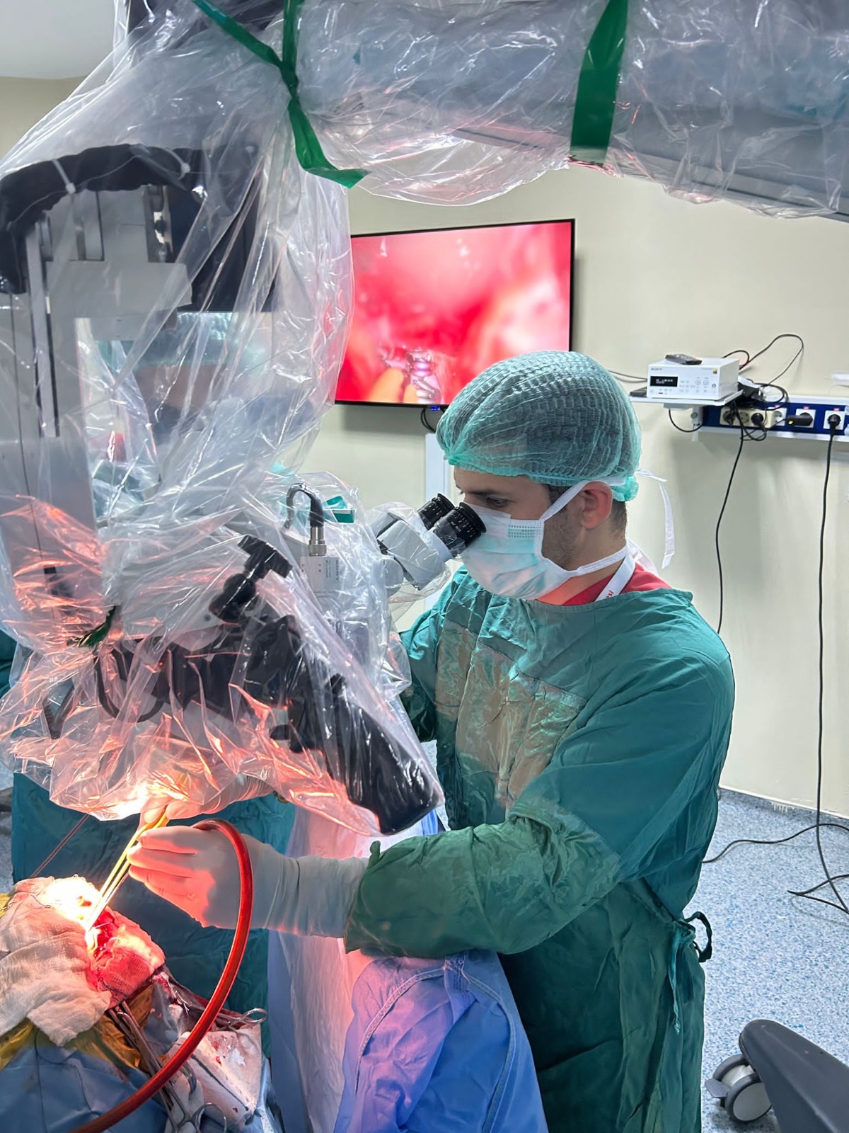
Operative Technique
Under general endotracheal anesthesia, patients’ heads were
positioned as turned 90° same side of the lesion so that the midline
was parallel to the floor (lateral neck flexion) and lifted 45° upward
(Figure 3A, Videos 1 and 2). A 3-pronged Mayfield skull clamp was
used for head fixation (Figure 3A). This approach allows for
gravitational retraction of the involved frontal or parietal lobe. The
incision site is shaved and prepared with a povidone-iodine solution.
After transcranial visualization of the lesion by navigation (Med-
tronic), the C shape skin incision, and craniotomy were performed
in accordance with the exact position of the lesion (Figure 3B). At
this stage, 2 burr holes are opened on the superior sagittal sinus (SSS)
and 2 more burr holes approximately 4 cm lateral to them (Videos 1
and 2). After performing the craniotomy, over the dura mater, the
navigation system was used to localize the central sulcus, precentral
and postcentral gyrus, as well as the trajectory of the lesion. The
location of the SSS is determined using a Doppler ultrasound (VTI
Vascular Technology, Inc., 20 MHz Doppler). The dura is opened
in a semicircular fashion with the base toward the SSS. Cortical veins
which drain into SSS are preserved (Figure 4A, Videos 1 and 2). The
dural flap is tacked upward to create a clear path along the falx
(Figure 4B, Videos 1 and 2). The arachnoid adhesions and
granulations adjacent to the SSS are removed with a micro-
surgical method, allowing gravity to pull the hemisphere away
from the falx and open the interhemispheric fissure without the
use of a retractor. The interhemispheric fissure is then opened
until the corpus callosum is encountered. The callosal sulcus is
observed above the corpus callosum, and the cingulate sulcus is
observed above the cingulate gyrus (Figure 4C, Videos 1 and 2).
The cingulate sulcus is dissected using a preserving cottonoid
FIGURE 3. A, With the ITCSA, the head is turned laterally 90° and neck is angled upward 45°. B, The intraoperative view down the right side of
the falx demonstrates the exposure of cingulate sulcus. Access to lesions in the deep-seated medial frontal lobe and parietal lobe with the ITCSA requires
an incision in the cingulate sulcus. ITCSA, interhemispheric transcingulate sulcus approach.
OPERATIVE NEUROSURGERY VOLUME 24 | NUMBER 3 | MARCH 2023 | E181
INTERHEMISPHERIC TRANSCINGULATE SULCUS APPROACH
© Congress of Neurological Surgeons 2023. Unauthorized reproduction of this article is prohibited.
Downloaded from http://journals.lww.com/onsonline by BhDMf5ePHKav1zEoum1tQfN4a+kJLhEZgbsIHo4XMi0hCyw
CX1AWnYQp/IlQrHD3i3D0OdRyi7TvSFl4Cf3VC1y0abggQZXdgGj2MwlZLeI= on 07/04/2023
placing (Figure 4D, Videos 1 and 2). Location of the lesion
relative to the cingular sulcus is determined by ultrasound (BK
Prosound α10). Ultrasound sonography imaging guides the
opening site. Bipolar forceps and microscissors were used as
much as feasible to dissect the sulcus. The vessels located in the
sulcus are preserved. Ultrasound sonography imaging confirms
complete removal of the lesion. This natural route in the brain
enables lesser injury in the crucial structures (Figure 4E,
Videos 1 and 2).
The operative steps, positioning, incision, and craniotomy for
this method are presented in attached videos (Videos 1 and 2).
Case 1
A 56-year-old male patient applied to our outpatient clinic
with complaints of headache, nausea, and weakness in left
extremities in April 2020. Computed tomography (CT) scan
revealed a round, high-density mass lesion in the right deep
white matter of the medial frontal lobe. Gadolinium (Gd)–
enhanced MRI showed a heterogeneous enhancement of the
tumor which settled deep in the medial frontal lobe just su-
perior to the cingulate gyrus, 14 × 19 × 22 mm in size (Figure
5A-5C). The tumor was completely removed using ITCSA
(Video 1). Postoperative MRI confirmed gross total resection
(Figure 5D-5F). Patient’s postoperative course was uneventful.
Pathology result was glioblastoma IDH wild type (WHO grade 4).
The preoperative extremity motor weakness decreased significantly
after the surgery.
Case 2
A 13-year-old boy reported a moderate headache in January
- He has a history of previous surgeries for temporal and
occipital cavernous malformations. Neurological examination was
unremarkable. A CT scan revealed a cavernoma in the deep white
matter of the medial frontal lobe. Gd-enhanced MRI showed a
heterogeneous enhancement of the cavernoma which located su-
perior to the cingulate sulcus and anterior to the marginal ramus,
15 × 11 × 17 mm in diameter (Figure 6A-6C). The cavernoma was
removed using ITCSA (Video 2). Postoperative MRI confirmed
total resection (Figure 6D-6F). Patient’s postoperative course
was uneventful. The preoperative headache resolved after the
procedure.
Case 3
A 54-year-old female patient applied to our outpatient clinic
with complaints of motor loss in left lower extremity and focal
epileptic seizure in November 2021. Gd-enhanced MRI revealed a
25 × 20-mm mass with heterogeneous contrast enhancement lo-
cated in the right precuneus at the level of the centrum semiovale
plane which was located posterior to the marginal ramus (Figure
7A-7C). The lesion was containing amorphous calcifications in the
central part (which was verified with CT scan), and perfusion MRI
showed increased perfusion. Radiological workup was compatible
with oligodendroglioma. The tumor was completely removed by
using ITCSA. Postoperative MRI and CT confirmed gross total
resection (Figure 7D-7F). Patient’s postoperative course was
FIGURE 4. Intraoperative view of the interhemispheric transcingulate sulcus approach. A, Cortical veins which drain into superior sagittal sinus are preserved. B, The dural flap is
tacked upward to create a clear path along the falx. C, The callosal sulcus is observed above the corpus callosum, and the cingulate sulcus is observed above the cingulate gyrus. D, The
cingulate sulcus is opened, and the cottonoid is placed at its entrance. E, The natural sulcus of the brain is used so that the structures are less damaged.
E182 | VOLUME 24 | NUMBER 3 | MARCH 2023 operativeneurosurgery-online.com
GUNGOR ET AL
© Congress of Neurological Surgeons 2023. Unauthorized reproduction of this article is prohibited.
Downloaded from http://journals.lww.com/onsonline by BhDMf5ePHKav1zEoum1tQfN4a+kJLhEZgbsIHo4XMi0hCyw
CX1AWnYQp/IlQrHD3i3D0OdRyi7TvSFl4Cf3VC1y0abggQZXdgGj2MwlZLeI= on 07/04/2023
uneventful. Pathology result was oligodendroglioma. The preop-
erative extremity motor weakness decreased significantly after the
surgery.
DISCUSSION
Lesions seated in the deep medial frontal and parietal lobes have
been one of the most challenging issues in neurosurgery because
of morbidities that may arise from damage to critical midline
structures or intact neural tissue that need to be crossed to reach
the lesion. Over the past 3 decades, several effective surgical
methods have been demonstrated for these lesions.1-11 The lateral
transcortical approach is still used by some surgeons18 which allows
more direct access to this area. The major disadvantage of this
approach is the significant injury of normal cortical tissue and
subcortical white matter during access19 and correlated increased
risk of morbidity. Neuromotor deficits that may arise from injury to
the motor cortex can be given as an example of these morbidities.19
Transcortical techniques can be performed with modest craniot-
omies and a direct path to the lesion, but they create a long and
unnatural surgical corridor through normal brain tissue.20,21
Despite having its own specific complications such as bridging
vein and dural sinus injury, medial approach to the hemisphere
has attracted the attention of neurosurgeons because it causes
less cortical damage. Ture et al, Spetzler et al, and Lawton et al
published several case series using the interhemispheric ap-
proach.7,9,16,19,22-24 Many different surgeries (transcallosal,
transcingulate, transchoroidal, and transrostral) were described in
these reports.
FIGURE 5. Patient 1: Preoperative magnetic resonance images demonstrating a high-grade glioma which settles in the deep medial frontal lobe, just superior to the cingulate
gyrus. A, Axial T1-weighted image with Gd; B, coronal T1-weighted image with Gd; and C, sagittal T1-weighted image with Gd images. Postoperative magnetic resonance
images demonstrated complete resection of the high-grade glioma. D, Axial T1-weighted image with Gd; E, coronal T1-weighted image with Gd; and F, sagittal T1-weighted
image with Gd images. Gd, gadolinium.
OPERATIVE NEUROSURGERY VOLUME 24 | NUMBER 3 | MARCH 2023 | E183
INTERHEMISPHERIC TRANSCINGULATE SULCUS APPROACH
© Congress of Neurological Surgeons 2023. Unauthorized reproduction of this article is prohibited.
Downloaded from http://journals.lww.com/onsonline by BhDMf5ePHKav1zEoum1tQfN4a+kJLhEZgbsIHo4XMi0hCyw
CX1AWnYQp/IlQrHD3i3D0OdRyi7TvSFl4Cf3VC1y0abggQZXdgGj2MwlZLeI= on 07/04/2023
During our anatomic studies, we found that the cingulate sulcus
could be an alternative route to access deep-seated lesions of the
medial frontal and parietal lobe, and we described the inter-
hemispheric transcingulate sulcus approach (ITCSA). Cingulate
sulcus, one of the brain’s natural pathway, was used in this pro-
cedure. ITCSA takes advantage of the interhemispheric fissure and
cingulate sulcus to provide deep access with minimal resection of
normal brain tissue. Patients tolerate the removal of a small portion
of the cingulate fibers and callosal fibers well. Many various surgical
diseases, including tumors, cysts, cavernomas, and arteriovenous
malformations, can be resected with this approach.
Horizontal head position is the key feature of this approach.1,6,8,9
Gravity enables withdraw of the involved hemisphere and modest
dissection of the interhemispheric fissure without further retraction
with blades in a horizontal head position with the lesion positioned
downside. Furthermore, this method is favorable because it makes
use of a natural sulcus of the cerebrum, allows for a more com-
fortable surgeon position, and provides a better trajectory. Trans-
sulcal methods reduce the length of the corridor through the brain
tissue, but they expose arteries and veins within the sulcus to
damage.7 ITCSA, in contrast to transcortical procedures, requires a
larger craniotomy, has a comparable working distance to the lesion,
but uses surgical corridor with many anatomic landmarks and the
brain’s natural sulcus.
In addition, the contralateral interhemispheric approach is also
used as an option. Lawton et al showed the advantages of the
contralateral approach.10 Even with gravity retraction, the ipsi-
lateral corridor is confined by the SSS and the medial surface of the
FIGURE 6. Patient 2: Preoperative magnetic resonance images demonstrated a cavernous malformation which settles deep in the medial frontal lobe, just superior to the cingulate
sulcus and anterior to the marginal ramus. A, Axial T2-weighted image; B, white matter tractography allowed to assess the relationships between the cavernoma and eloquent
tracts, coronal image; C, sagittal T2-weighted images; D, postoperative magnetic resonance images demonstrated complete resection of the cavernous malformation on axial T2-
weighted; E, coronal T2-weighted images; and F, sagittal T2-weighted images.
E184 | VOLUME 24 | NUMBER 3 | MARCH 2023 operativeneurosurgery-online.com
GUNGOR ET AL
© Congress of Neurological Surgeons 2023. Unauthorized reproduction of this article is prohibited.
Downloaded from http://journals.lww.com/onsonline by BhDMf5ePHKav1zEoum1tQfN4a+kJLhEZgbsIHo4XMi0hCyw
CX1AWnYQp/IlQrHD3i3D0OdRyi7TvSFl4Cf3VC1y0abggQZXdgGj2MwlZLeI= on 07/04/2023
brain, resulting in slightly shorter working distances than with the
contralateral transcingulate approach. This narrower corridor is
beneficial for lesions on the surface of the cingulate gyrus, but it is
unfavorable for lesions that extend laterally into the deep frontal
lobe. The contralateral transcingulate approach can be used as an
alternative for these deep frontal lesions, providing direct view of
the lesion as well as easier mobility because of the crossing tra-
jectory through the interhemispheric fissure.
Our surgical experience has shown that the ITCSA is an al-
ternative way to treat lesions located deep in the medial frontal
lobe and parietal lobe without causing any neurological deficits.
We believe that in most cases, total surgical resection of these
lesions is possible when supported by extensive anatomic
knowledge and enhanced microsurgical technique.
Limitations
Due to the small number of cases in our series, the advantages
and disadvantages of the approach can be better demonstrated by
conducting extensive studies. In addition, ITCSA has limitations
similar to the interhemispheric approach, such as bridging vessel
and dural sinus injury.
CONCLUSION
Our anatomic study and cases show that the ITCSA is an
alternative way to reach deep-seated medial frontal lobe and
parietal lobe lesions causing less damage to white matter tracts and
preserving eloquent cortical areas.
FIGURE 7. Patient 3: Preoperative magnetic resonance images demonstrating a heterogeneously contrast-enhanced mass located in the right precuneus in the centrum semiovale
plane. A, Sagittal DTI-MR tractography; B, coronal DTI-MR tractography; and C, sagittal T1-weighted image with gadolinium images. Postoperative magnetic resonance and
computed tomography images demonstrated complete resection of the high-grade glioma. D, Axial CT image; E, coronal CT; and F, sagittal T1-weighted image with gadolinium
images. CT, computed tomography; DTI-MR, diffusion tensor imaging magnetic resonance.
OPERATIVE NEUROSURGERY VOLUME 24 | NUMBER 3 | MARCH 2023 | E185
INTERHEMISPHERIC TRANSCINGULATE SULCUS APPROACH
© Congress of Neurological Surgeons 2023. Unauthorized reproduction of this article is prohibited.
Downloaded from http://journals.lww.com/onsonline by BhDMf5ePHKav1zEoum1tQfN4a+kJLhEZgbsIHo4XMi0hCyw
CX1AWnYQp/IlQrHD3i3D0OdRyi7TvSFl4Cf3VC1y0abggQZXdgGj2MwlZLeI= on 07/04/2023
Funding
This study did not receive any funding or financial support.
Disclosures
The authors have no personal, financial, or institutional interest in any of the
drugs, materials, or devices described in this article.
REFERENCES
- Benet A, Griswold D, Tabani H, et al. Contralateral anterior interhemispheric
approach to medial frontal arteriovenous malformation: 3-dimensional operative
video. Oper Neurosurg. 2018;14(1):86-86. - Chaddad-Neto F, Joaquim AF, dos Santos MJ, Linhares PW, de Oliveira E.
Microsurgical approach of arteriovenous malformations in the central lobule. Arq
Neuropsiquiatr. 2008;66(4):872-875. - Fernandez-Miranda JC, Xu Y, Hendricks BK, Cohen-Gadol A. Contralateral
Interhemispheric transfalcine transprecuneus approach: advancing operative angles
to deep-seated lesions. World Neurosurg. 2020;144:341-350. - Glauser G, Abdullah KG, Choudhri OA. Contralateral transcingulate resection of
midcingulate cavernous malformation. World Neurosurg. 2019;129:389. - Tominaga T, Kayama T, Kumabe T, Yoshimoto T. Transcingulate approach to
lateral ventricle tumors. Technical case report. Neurosurg Rev. 1996;19(2):105-108. - Burkhardt JK, Winkler EA, Lawton MT. Contralateral posterior interhemispheric
approach to deep medial parietooccipital vascular malformations: surgical technique
and results. J Neurosurg. 2018;129(1):198-204. - Mikuni N, Hashimoto N. A minimally invasive transsulcal approach to the
paracentral inner lesion. Minim Invasive Neurosurg. 2006;49(5):291-295. - Zaidi HA, Chowdhry SA, Nakaji P, Abla AA, Spetzler RF. Contralateral inter-
hemispheric approach to deep-seated cavernous malformations: surgical consider-
ations and clinical outcomes in 31 consecutive cases. Neurosurgery. 2014;75(1):80-86. - Chaddad-Neto F, Devanir Silva da Costa M, Bozkurt B, et al. Contralateral anterior
interhemispheric-transcallosal-transrostral approach to the subcallosal region: a
novel surgical technique. J Neurosurg. 2018;129(2):508-514. - Davies J, Tawk RG, Lawton MT. The contralateral transcingulate approach:
operative technique and results with vascular lesions. Oper Neurosurg. 2012;71(1
suppl operative):ONS4-ONS14. - Yano S, Hide T, Shinojima N, Ueda Y, Kuratsu JI. A flexible endoscope-assisted
interhemispheric transcallosal approach through the contralateral ventricle for the
removal of a third ventricle craniopharyngioma: a technical report. Surg Neurol Int.
2015;6(suppl 2):S113-S116. - Klingler J. Erleichterung Der Makrokopischen Pr ̈aparation Des Gehirns Durch Den
Gefrierprozess. Orell Füssli; 1935. - Ribas GC. The cerebral sulci and gyri. Neurosurg Focus. 2010;28(2):E2.
- Güng ̈or A, Baydin S, Middlebrooks EH, Tanriover N, Isler C, Rhoton AL. The
white matter tracts of the cerebrum in ventricular surgery and hydrocephalus.
J Neurosurg. 2017;126(3):945-971. - Baydin S, Gungor A, Tanriover N, Baran O, Middlebrooks EH, Rhoton AL. Fiber tracts
of the medial and inferior surfaces of the cerebrum. World Neurosurg. 2017;98:34-49. - Panteli A, Güng ̈or A, Fırat Z, Sarıtepe F, Türe H, Türe U. The posterior inter-
hemispheric transparieto-occipital fissure approach to the atrium of the lateral ventricle: a
fiber microdissection study with case series. Neurosurg Rev. 2022;45(2):1663-1674.
- Gurses ME, Gungor A, Hanalioglu S, et al. Qlone®: a simple method to create 360-
degree photogrammetry-based 3-dimensional model of cadaveric specimens. Oper
Neurosurg. 2021;21(6):E488-E493. - Hameed NUF, Wu B, Gong F, Zhang J, Chen H, Wu J. Awake transcortical
approach resection of dominant posterior cingulate gyrus glioma: 2-dimensional
operative video. Oper Neurosurg. 2019;17(1):E19-E20. - Abla AA, Spetzler RF, Albuquerque FC. Trans-striatocapsular contralateral in-
terhemispheric resection of anterior inferior basal ganglia cavernous malformation.
World Neurosurg. 2013;80(6):e397-e399.
- Fehlings MG, Houlden D, Vajkoczy P. Introduction. Intraoperative neuro-
monitoring: an essential component of the neurosurgical and spinal armamen-
tarium. Neurosurg Focus. 2009;27(4):E1. - Sala F, Krzan MJ, Deletis V. Intraoperative neurophysiological monitoring in pe- ˇ
diatric neurosurgery: why, when, how? Child’s Nervous Syst. 2002;18(6-7):264-287. - Serra C, Türe U. The extreme anterior interhemispheric transcallosal approach for
pure aqueduct tumors: surgical technique and case series. Neurosurg Rev. 2022;
45(1):499-505. - Hendricks BK, Spetzler RF. Contralateral interhemispheric transcallosal trans-
choroidal approach for resection of a tumor in the lateral wall of the third ventricle:
2-dimensional operative video. Oper Neurosurg. 2019;17(5):E197.
- Hendricks BK, Spetzler RF. Contralateral interhemispheric approach for resection of a
meningioma: 2-dimensional operative video. Oper Neurosurg. 2020;18(5):E159.
VIDEO 1. Case 1.
VIDEO 2. Case 2.
COMMENT
Mankind has a long interest for studying the brain, but interestingly,
it was only in the middle of the 19th century that the anatomic
organization of the cerebral sulci and gyri was perceived and detailed, with
great contributions from French anatomists. At that time, Achille Louis
Foville provided an atlas with accurate drawings of the brain surface, followed
by Louis Pierre Gratiolet, known for the unprecedented recognition of a
regular arrangement of cerebral sulci and gyri among all human brains despite
individual variations. Paul Broca noted the peculiar C shape of the cingulate
gyrus continuing posteriorly and inferiorly to the parahippocampal gyrus,
having the diencephalon at the center, and named them as the greater limbic
lobe. He also considered the cingulate, subparietal, and collateral sulci to be
segments of the sulcus he referred to as the limbic sulcus.
The development of microneurosurgery, together with deeper
knowledge about the cortical surface and the subcortical white matter
tracts organization, made the use of cerebral sulci as microneurosurgical
corridors possible. Several transcisternal, transfissural, and transsulcal
approaches have been established to approach subcortical and intra-
ventricular lesions, particular through the sulci of the superolateral and
inferior surfaces of the brain, which are consistently oriented toward the
nearest ventricular cavity because are formed by mechanisms of cortical
invagination throughout evolution and embryology. At the medial
surface of the brain, this sulci disposition is not seen because their de-
velopment is directly related to that of the corpus callosum, and these
sulci therefore tend to be arranged in parallel with this commissure.
The medial surface of the brain is not exposed prima facie during
surgeries. It requires broad and meticulous dissection of the interhemi-
spheric fissure to allow retraction of the medial surface and delicate work
between bridging veins to the superior sagittal sinus. The authors incor-
porated these concepts and, with great microsurgical skill, demonstrated
that the cingulate sulcus can also be used as a safe surgical corridor to deep-
seated lesions at frontal and parietal lobes. Congratulations.
Eduardo Carvalhal Ribas
Guilherme Carvalhal Ribas
São Paulo, Brazil