ABSTRACT
Objective: To analyse the relationship between peritumoural oedema volume and tumour volume in relation to the impact of
metastatic posterior fossa tumour survival rates.
Study Design: An observational study.
Place and Duration of Study: Umraniye Training and Research Hospital, İstanbul, Turkey, from 2011–2021.
Methodology: Fifty-six cancer patients who had been operated upon for cerebellar metastases were analysed retrospectively. To
investigate the effect of oedema on survival, patients with a single cerebellar metastasis were evaluated retrospectively. Those patients
had a single metastasis located in the cerebellum and did not receive radiotherapy or corticosteroids before surgery. OsiriX MD DICOM

viewer was used to calculate the volumes of the tumour and the oedema using fluid-attenuated inversion recovery (FLAIR) and contrast-
enhanced magnetic resonance imaging (MRI). The patients were separated into two groups, and the cut-off limit for the oedema to-tu-
mour ratio was set to two. Survival analysis was performed on the two groups.

Results: When the primary sites of the tumours were evaluated, 60.7% were located in the lungs (n = 34), 10.7% were located in the
breasts (n = 6), 10.7% were located in the gastrointestinal tract (n = 6), 7.1% were located in the renal region (n = 4), 5.4% were
located in the gynaecologic tract (n = 3), and 5.4% were located in other parts of the body (n = 3). A univariate analysis showed that
overall survival duration was significantly longer in the subgroup with breast cancer (83.3%) and in those patients with a peritumoural

oedema volume to tumour volume ratio of less than two (27.6%, p <0.05). Negative prognostic factors were lung cancer and high peritu-
moural oedema volume.

Conclusion: Significant peritumoural oedema was linked to a poor prognosis for cancer patients with a single cerebellar metastasis,
especially with lung cancer as the primary source.
Key Words: Cerebellar metastases, Cerebellum, OsiriX MD, Tumour volume.
How to cite this article: Ramazanoglu AF, Varol E, Avci F, Etli MU, Aydin S, Yaltirik CK. Peritumoural Oedema as a Predictor of Overall
Survival for Patients with Posterior Fossa Metastases. J Coll Physicians Surg Pak 2023; 33(02):136-140.

INTRODUCTION

Intracranial metastases are the common complication in cancer

patients, and 30–40% of cancer patients develop brain metas-
tases at some point during the course of their disease.1

A total of
20% of brain metastases occur in the posterior fossa.2
If the

metastasis remains untreated, the patient runs the risk of devel-
oping serious conditions such as acute hydrocephalus, brain-
stem compression, cerebellar tonsillar herniation, and falling

into a coma.2

There are various factors that affect the overall

survival rates for intracranial metastases patients,3,4 with func-
tional impairment (measured using the Karnofsky Performance

Scale Index), the occurrence of extracranial metastases, and
adjuvant chemotherapy or radiotherapy being the primary
factors that affect life expectancy.1,5,6

Correspondence to: Dr. Cumhur Kaan Yaltirik, Depart-
ment of Neurosurgery, Umraniye Training and Research

Hospital, Istanbul, Turkey
E-mail: dr_cky@yahoo.com
……………………………………………..
Received: July 30, 2022; Revised: December 11, 2022;
Accepted: January 03, 2023
DOI: https://doi.org/10.29271/jcpsp.2023.02.136

However, there are only a few studies in literature on the radiolog-
ical factors impacting the estimated survival rates of patients

with posterior fossa metastasis.7

In this study, the aim was to find
the correlation between peritumoural oedema volume and
tumour volume in relation to the impact of metastatic posterior
fossa tumours on patient survival rate.
In this study, 172 patients who had been operated on at the
Ümraniye Research and Training Hospital, between 2011 and
2021 were studied retrospectively using their hospital records.
To investigate the effect of oedema on survival, patients with a
single cerebellar metastasis were evaluated retrospectively.
The patients had a single metastasis located in the cerebellum
and did not receive radiotherapy or corticosteroids before
surgery. The pathology report was compatible with metastasis,
and the preoperative magnetic resonance imaging (MRI)
included both contrast-enhanced images and fluid-attenuated
inversion recovery (FLAIR) sequences, which were included in
the study. Patients who did not have a brain MRI prior to surgery,

whose clinical information could not be obtained, who had multi-
focal cerebral or cerebellar metastasis, who did not have a

primary focus of cancer, who were diagnosed with a neurodegen-
erative disease, and who received corticosteroid therapy for any

reason were excluded from the study. Following these criteria,

Ali Fatih Ramazanoglu, Eyup Varol, Furkan Avci, Mustafa Umut Etli, Serdar Aydin and Cumhur Kaan Yaltirik

Journal of the College of Physicians and Surgeons Pakistan 2023, Vol. 33(02): 136-140 137
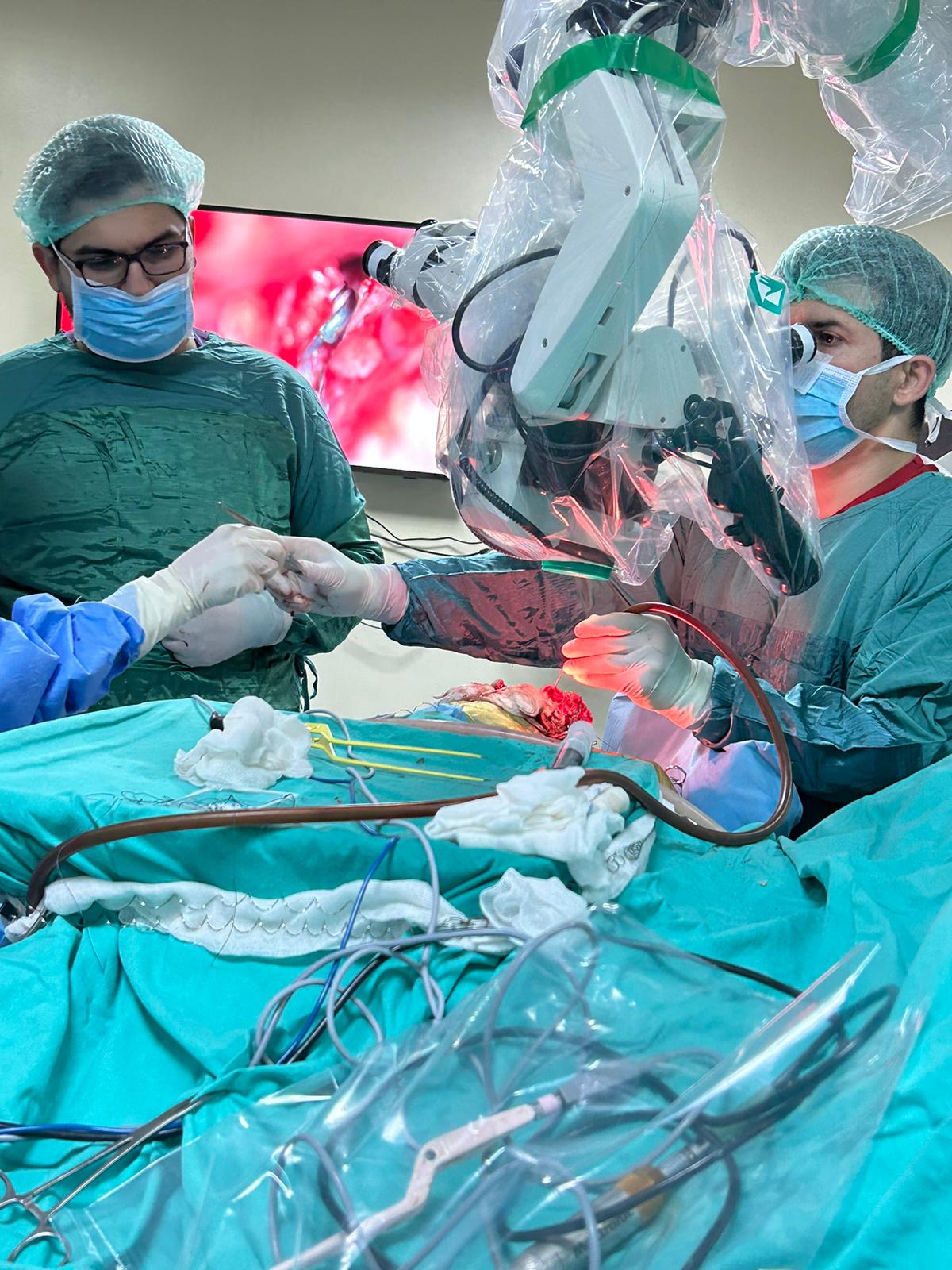
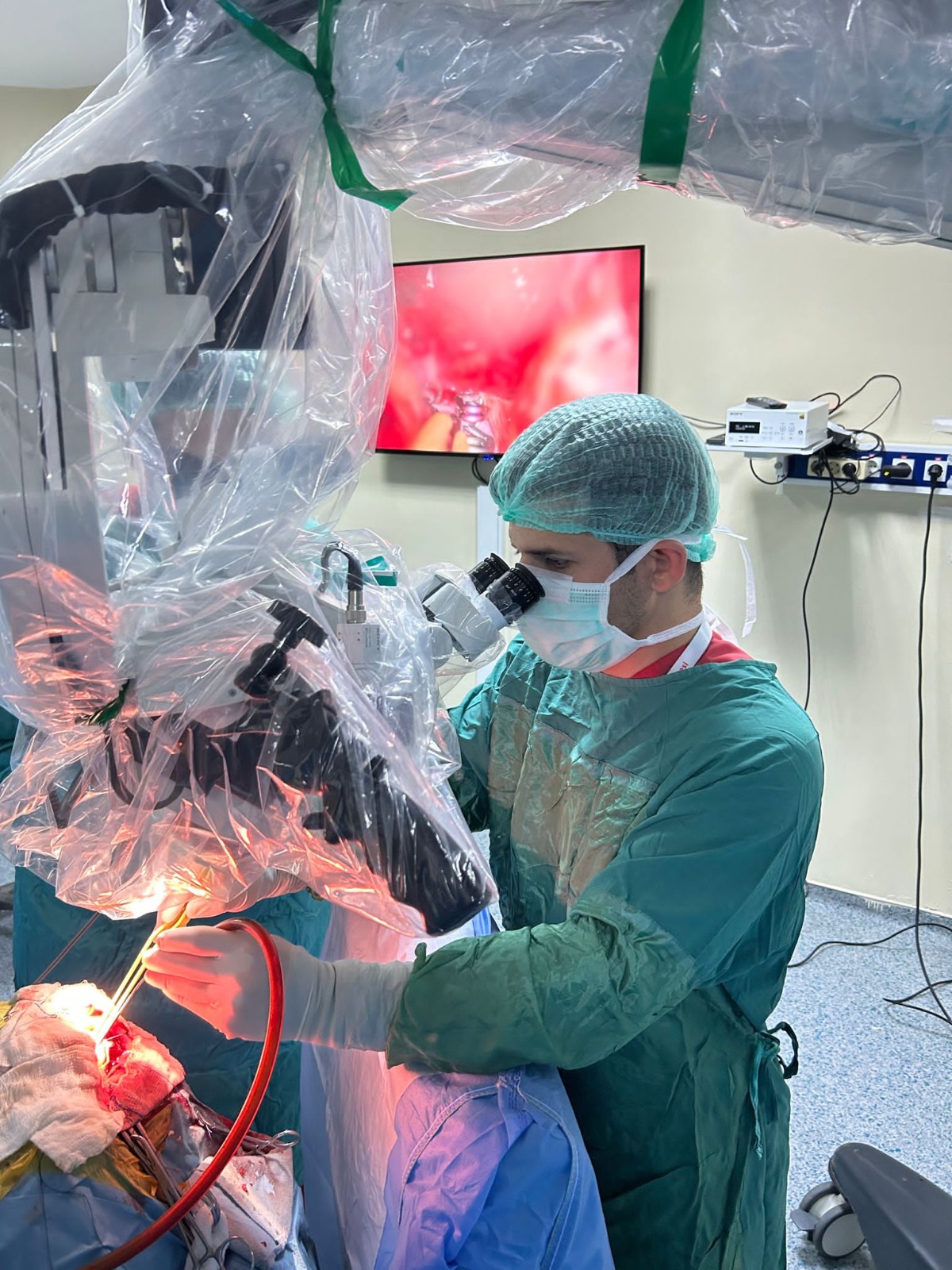
56 patients were retrospectively analysed. All study participants
had undergone surgical resection and received radiotherapy
during the postoperative period. Thirty-four patients with a score
of less than two and twenty-two patients with a score of more

than two were included in the study. The cut-off limit for the oede-
ma-to-tumour ratio was two. The participants were divided into

two groups, and survival analysis was performed on two groups.
OsiriX MD is cleared by the FDA as a class II medical device for
diagnostic imaging. Hence, OsiriX MD software was used to
calculate tumour volume and peritumoural oedema volume.8
The tumours were viewed on T1 contrast-enhanced MRI
images, and a region of interest9

was marked around the tumour
tissue. Peritumoural oedemas were viewed via MRI, ROIs, and
FLAIR sequences were marked around the peritumoural
oedema tissue. The markings were made on every section of the

MRI images, and the peritumoural oedema volume was calcu-
lated using OsiriX software. To isolate the peritumoural oedema

volume from the tumour volume, the tumour volume was
subtracted from the total volume, and the peritumoural volume
was obtained. The ratio of peritumoural oedema volume to
tumour volume was then calculated (Figures 1a-1c). The
measurements were performed by two different researchers

simultaneously; there were no statistically significant interob-
server differences between the measurements.

Figure 1 (a,b,c): Calculation of tumour volume and peritumoural oedema
volume using OsiriX MD. (d,e): Kaplan–Meier survival analysis of the
study groups.
Statistical Package for the Social Sciences (SPSS) version 25.0

(IBM Corp., Armonk, NY, USA) was used for the statistical anal-
ysis of the results. Descriptive statistics, including numbers,

percentages, and mean and standard deviation (SD) were used
to evaluate the results of the study. The Kaplan–Meier analysis
method was used to calculate survival. The effects of prognostic

factors on survival were evaluated using Cox regression anal-
ysis. The results were evaluated with 95% confidence interval

and p <.05 was considered significant.
RESULTS

The median age of the participants was 62 (30–87), and 50%
were 63 years or older (n = 28). A total of 69.6% (n = 39) of the

patients were male, and 30.4% (n = 17) were female. When the
primary sites of the cancer tumours were evaluated, 60.7%
were located in the lungs (n = 34), 10.7% were located in the
breasts (n = 6), 10.7% were located in the gastrointestinal tract
(n = 6), 7.1% were located in the renal region (n = 4), 5.4% were
located in the gynaecologic tract (n = 3), and 5.4% were located
in other areas (n = 3). The median volume of the tumours
viewed in T1 contrast-enhanced MRI images was 9.54 cm3
(0.28–30.1 cm3), and the median volume of the peritumoural
oedemas, which was calculated using the total peritumoural
oedema volume in FLAIR sequences—was 92.99 cm3
(11.99–130.85 cm3). The peritumoural oedema volume to
tumour volume was less than two in 60.7% of the participants (n
= 34) and greater than two in 39.3% (n = 22, Table I).
Twenty-two of the 56 study participants (39.2%) had systemic
metastases, four (18.18%) had adrenal gland metastases, four
(18.18%) had lung metastases, and four (18.18%) had spinal
distant metastases. Hydrocephalus was observed in 16 (29%)
participants, while only four (7.1%) participants became

ventriculoperitoneal shunt-dependent. Twelve study partici-
pants (75%) recovered from hydrocephalus after being treated

with an external ventricular drainage system.

During follow-up, 73.2% of the participants (n = 41) experi-
enced death related to intracranial tumours (mean: 14.14 ±

24.62 months, median: 4.3 (1–124) months), and 60.7% of the
deaths occurred within a year. The one and three-year survival
rates were 36.7% and 21.9%, respectively, based on a
Kaplan–Meier survival analysis (Figure 1d and 1e). A univariate
analysis showed that overall survival was significantly longer in
the breast cancer subgroups, and the peritumoural oedema
volume to tumour volume ratio was less than two (27.6%, p <
0.05). It was found that the negative prognostic factors
impacting overall survival were of lung cancer [HR: 7.71
(1.04–57.32), p = 0.046], and the presence of other metastatic
tumours [HR: 11.56 (1.51–88.57), p = 0.018]. Based on the

results of the multivariate Cox regression analysis, the indepen-
dent factors affecting overall survival were lung cancer [HR:

12.98 (1.51–111.64), p = .020], multiple cancer diagnoses [HR:
10.27 (1.23–86.04), p = .032], and an oedema to tumour ratio
greater than two [HR: 2.13 (1.01–4.52), p = .048].
Compared to breast cancer patients as a reference, overall
survival was 12.98 times more negative in lung cancer
patients, 10.27 times more negative in patients with multiple
cancer diagnoses, and 2.13 times more negative in patients
whose peritumoural oedema volume to tumour volume ratio
was greater than two (Table II).
DISCUSSION

Davis et al. found that the most common primary site of malig-
nancy for brain metastases are the lungs (20–40%).9

However,
60.7% of the posterior fossa metastases in this study originated

from a lung. The lung cancer metastases in this study is signifi-
cantly greater than in other studies.10

Peritumoural oedema as a predictor of overall survival with posterior fossa metastases

138 Journal of the College of Physicians and Surgeons Pakistan 2023, Vol. 33(02): 136-140
Table I: Demographic Information (n = 56).
Variable N % Mean (SD) Median (range)
Age 56 100 60.59 (11.11) 62.5 (30–87)
Age group
<63 28 50
≥63 28 50
Gender
Female 17 30.4
Male 39 69.6
Tumour localisation
Lung 34 60.7
Breast 6 10.7
Gastrointestinal tract 6 10.7
Renal region 4 7.1
Gynaecologic tract 3 5.4
Other 3 5.4
Tumour volume (cm3) 56 100 9.83 (6.50) 9.54 (0.28–30.1)
Peritumoural oedema
volume (cm3)

56 100 16.42 (12.82) 16.57
(0.14–70.48)

Ratio of peritumoural
oedema volume to
Tumour volume

56 100 3.22 (3.77) 1.71 (0.01–16.85)

Ratio of peritumoural
oedema volume to tumour
volume
≤2 34 60.7

2 22 39.3
Systemic metastasis
Yes 22 39.3
No 34 60.7
Systemic metastasis type
Adrenal gland metastases 4 18.2
Lung metastases 4 18.2
Spinal distant metastases 4 18.2
Other 10 45.5
Hydrocephalus
complication
Yes 16 28.6
No 40 71.4
Ventriculoperitoneal shunt
Yes 4 7.1
No 52 92.9
SD: Standard deviation.

Magnetic resonance (MR) examinations, which were adopted
for clinical use in the 1980s, have taken the place of computed
tomography (CT) examinations in diagnosing primary brain
tumours and evaluating patient response to treatment.11
Contrast-enhanced MRI, which is widely used in metastasis
screening, facilitates detailed evaluation of parenchymal
metastases and structures such as the calvarium, diploic
distance, meninges, choroid plexus, and pituitary gland.11

FLAIR sequences suppress the T2-hyperintensity of cere-
brospinal fluid surrounding the brain parenchyma and in the

ventricles, permitting a clearer assessment of oedema adja-
cent to the lesion.12 MRI has a higher sensitivity than CT scans

and positron emission tomography PET-CT scans for detecting
small metastases.13,14

Advanced MRI imaging methods are frequently used in metas-
tasis examinations.15 MRI perfusion examination provides

information on the blood flow to the lesion.9

In this study,
volume measurements performed on acoustic neuromas by
Kollman et al. using Brainlab software, ITK-Snap, and the OsiriX
radiological DICOM viewer system were tested on acoustic
neuromas.16 OsiriX MD has been shown to be faster and more
reliable for volume measurements than other systems.16

The main purpose of this study is to understand whether peritu-
moural oedema of posterior fossa metastatic tumours impacts

the overall survival of cancer patients. The peritumoural
oedema volume to tumour volume ratio has recently been
investigated for diagnostic purposes by other researchers. The
literature review revealed that this study is the third research
evaluating survival in cerebellar metastasis patients. Calluaud
et al. found that peritumoural oedema is highly significant to
the overall survival of patients with metastatic posterior fossa
tumours.17 The authors divided the patients in their study into
four groups based on peritumoural oedema volume to tumour
volume ratio and found that overall survival was significantly
low with a cut-off ratio of two—a result that is in line with
authors study. They also simultaneously investigated patients
with supratentorial metastasis and posterior fossa metastasis,

while the authors excluded patients with supratentorial metas-
tasis from the study.

Kaya et al. found that carcinoma metastases have larger peritu-
moural oedema volumes than non-carcinoma metastase;

overall. However, the survival rates reported were not signifi-
cantly different in that study.18 Here, only cerebellar metas-
tases and the effect of oedema around the tumour on the

survival of the patient were evaluated. Patients with a peritu-
moural oedema volume to tumour volume ratio greater than

two had significantly shorter overall survival than patients with
a lower ratio. The authors used OsiriX MD software for
measurements, and Kaya et al. and Calluaud et al. also
used the same OsiriX software to measure tumour and
metastasis volumes.17,18

Ali Fatih Ramazanoglu, Eyup Varol, Furkan Avci, Mustafa Umut Etli, Serdar Aydin and Cumhur Kaan Yaltirik

Journal of the College of Physicians and Surgeons Pakistan 2023, Vol. 33(02): 136-140 139
Table II: Factors affecting overall survival (univariate and multivariate Cox regression analysis results).

Univariate
overall survival analysis

Multivariate
overall survival analysis

Factor Category % HR (95%CI) p-value HR (95%CI) p-value
Age <63 33.9

≥63 0.07 1.77 (0.94–3.32) 0.077 1.33 (0.69–2.55) 0.395

Gender Female 35.3

Male 14.5 1.25 (0.62–2.51) 0.533 1.99 (0.87–4.55) 0.104

Tumour location Breast 83.3

Lung 12.8 7.71 (1.04–57.32) 0.046* 12.98 (1.51–111.64) 0.020*
Other 12.5 11.56 (1.51–88.57) 0.018* 10.27 (1.23–86.04) 0.032*

Ratio of peritumoural
oedema volume to
Tumour volume

≤2 27.6

2 13.9 2.07 (1.10–3.88) 0.023* 2.13 (1.01–4.52) 0.048*

*p <0.05, Cox regression analysis, (Method = Enter), HR: Hazard ratio, CI: Confidence interval.
Previous studies have reported that 8% of cancer patients
develop hydrocephalus.2,19 In this study, 28.5% of the
studied patients developed hydrocephalus (n = 16). Only

25% of the study participants who developed hydro-
cephalus required a ventriculoperitoneal shunt operation,

and 75% went through the perioperative period using an
external ventricular drainage catheter; there was no shunt
dependency.

Limitations of this study overall survival investigated in rela-
tion to only peritumoural oedema volume to tumour volume

and not adjuvant radiotherapy or chemotherapy. Moreover,
the impact of other known factors on overall survival was
not investigated, e.g., graded prognostic assessment (GPA);

resection rates, preoperative comorbidities such as hydro-
cephalus, and molecular variations were not considered in

the statistical analysis. In future studies, peritumoural
oedema should be examined together with these factors,
and prognostic algorithms should be created. The method

used in this study allowed the authors to find the correla-
tion between preoperative neuroimaging findings and

overall survival, revealing that a significant peritumoural
oedema, as indicated by a high oedema volume to tumour
volume ratio, is linked to a poor prognosis.
CONCLUSION

Significant peritumoural oedema is linked to a poor prog-
nosis for cancer patients with a single cerebellar metas-
tasis, particularly in patients with primary lung cancer.

ETHICAL APPROVAL:
The study was approved by the ethical committee of the
Umraniye Training and Research Hospital. (Reference No.
B.10.1. TKH.4.34.H.GP.0.01/221).
PATIENTS’ CONSENT:
Informed consents were obtained from all patients included
in the study.
COMPETING INTEREST:
The authors declare that there are no conflicts of interest
concerning the materials or methods used in this study or
the findings reported in this paper.
AUTHORS’ CONTRIBUTION:
AFR, MUE, CKY: Contributed to the conception and design of
the study.
AFR, EV, FA, MUE, SOA, CKY: Contributed to patient inclusion
and follow-up.

REFERENCES

  1. Kotecha R, Gondi V, Ahluwalia MS, Brastianos PK, Mehta MP.
    Recent advances in managing brain metastasis. F1000Res
    2018; 7. doi.org/10.12688/f1000research.15903.1.
  2. Sunderland GJ, Jenkinson MD, Zakaria R. Surgical manage-
    ment of posterior fossa metastases. J Neurooncol 2016;

130(3): 535-42. doi.org/10.1007/s11060-016-2254-2.

  1. Sivasanker M, Madhugiri VS, Moiyadi AV, Shetty P, Subi TS.
    Surgery for brain metastases: An analysis of outcomes and
    factors affecting survival. Clin Neurol Neurosurg 2018; 168:
    153-62. doi.org/10.1016/j.clineuro.2018.03.011.

Peritumoural oedema as a predictor of overall survival with posterior fossa metastases

140 Journal of the College of Physicians and Surgeons Pakistan 2023, Vol. 33(02): 136-140

  1. Xiao W, Li X, Yang A. Analysis of prognostic factors affecting
    the brain metastases free survival and survival after brain
    metastases in breast cancer. Front Oncol 2020; 10: 431.
    doi.org/10.3389/fonc.2020.00431.
  2. Liu Z, Lei B, Zheng M, Li Z, Huang S, Deng Y. Prognostic

factors in patients treated with surgery for brain metas-
tases: A single-center retrospective analysis of 125 patients.

Int J Surg 2017; 44: 204-9. doi.org/10.1016/j.ijsu.2017.05.
033.

  1. Rastogi K, Bhaskar S, Gupta S, Jain S, Singh D, Kumar P.
    Palliation of brain metastases: Analysis of prognostic factors
    affecting overall survival. Indian J Palliat Care 2018; 24(3):
    308-12. doi.org/10.4103/IJPC.IJPC_1_18.
  2. Yaltirik Bilgin E, Unal O, Ciledag N. Vasogenic oedema
    Pattern in Brain Metastasis. J Coll Physicians Surg Pak 2022;
    32(8):1020-5. doi.org/10.29271/jcpsp.2022.08.1020.
  3. Lovato RM, Araujo JLV, Paiva ALC. The use of osirix for surg-
    ical planning using cranial measures and region of interest

tools: Technical note. Asian J Neurosurg 2019; 14(3): 762-6.
doi.org/10.4103/ajns.AJNS_63_19.

  1. Essig M, Nguyen TB, Shiroishi MS. Perfusion MRI: The five
    most frequently asked clinical questions. AJR Am J

Roentgenol 2013; 201(3):W495-510. doi.org/10.2214/A-
JR.12. 9544.

  1. Popper HH. Progression and metastasis of lung cancer.
    Cancer Metastasis Rev 2016; 35(1): 75-91. doi.org/10.
    1007/s10555-016-9618-0.
  2. Maroldi R, Ambrosi C, Farina D. Metastatic disease of the
    brain: Extra-axial metastases (skull, dura, leptomeningeal)
    and tumour spread. Eur Radiol 2005; 15(3):617-26.
    doi.org/10.1007/s00330-004-2617-5.
  3. Smirniotopoulos JG, Murphy FM, Rushing EJ, Rees JH,
    Schroeder JW. Patterns of contrast enhancement in the

brain and meninges. Radiographics 2007; 27(2):525-51.
doi.org/10.1148/rg.272065155.

  1. Graif M, Bydder GM, Steiner RE, Niendorf P, Thomas DG,
    Young IR. Contrast-enhanced MR imaging of malignant brain
    tumours. AJNR Am J Neuroradiol 1985; 6(6): 855-62.
  2. Chen WS, Li JJ, Hong L, Xing ZB, Wang F, Li CQ. Comparison
    of MRI, CT and 18F-FDG PET/CT in the diagnosis of local and
    metastatic of nasopharyngeal carcinomas: An updated meta
    analysis of clinical studies. Am J Transl Res 2016; 8(11):
    4532-47.
  3. Chow KKH, Meola A, Chang SD. Commentary: Peritumoural
    oedema/tumour volume ratio: A strong survival predictor for
    posterior fossa metastases. Neurosurgery 2019; 85(1): E18-
    E19. doi.org/10.1093/neuros/nyy281.
  4. Kollmann P, Mautner VF, Koeppen J. MRI based volumetric
    measurements of vestibular schwannomas in patients with

neurofibromatosis type 2: Comparison of three different soft-
ware tools. Sci Rep 2020; 10(1): 11541. doi.org/10.1038/

s41598-020-68489-y.

  1. Calluaud G, Terrier LM, Mathon B. Peritumoural oedema/tu-
    mour volume ratio: A strong survival predictor for posterior

fossa metastases. Neurosurgery 2019; 85(1):117-25.
doi.org/10.1093/neuros/nyy222.

  1. Kaya I, Cingoz ID, Gursoy M. Oedema-mass ratio based on
    magnetic resonance imaging as a preoperative diagnostic
    factor for posterior fossa metastasis. Curr Med Imaging
    2021; 17(6): 762-6. doi.org/10.2174/157340561766621030
    3105006.
  2. Roux A, Botella C, Still M. Posterior fossa metastasis-associ-
    ated obstructive hydrocephalus in adult patients: Literature

review and practical considerations from the neuro-on-
cology club of the french society of neurosurgery. World

Neurosurg 2018; 117: 271-9. doi.org/10.1016/ j.wneu.2018.
06.084.

*Department of Neurosurgery, Ondokuz Mayis University, School of Medicine, Samsun, Turkey; ‡

Department of Neurosurgery, University of Health Sciences, Bakirkoy Prof. Dr.

Mazhar Osman Training and Research Hospital for Neurology, Neurosurgery and Psychiatry, Istanbul, Turkey; §

Department of Neurosurgery, Ankara University, School of Medicine,

Ankara, Turkey; ||Department of Neurosurgery, Hacettepe University, School of Medicine, Ankara, Turkey; ¶

Department of Neurosurgery, University of Health Sciences, Umraniye

Teaching and Research Hospital, ̇

Istanbul, Turkey; #

Department of Neurosurgery, General University Hospital Alicante, Alicante, Spain; **Department of Neurosurgery, Indiana

University School of Medicine, Indianapolis, Indiana, USA; ††The Neurosurgical Atlas, Carmel, Indianapolis, Indiana, USA; ‡‡Department of Neurosurgery, Microsurgical Neuro-
anatomy Laboratory, Yeditepe University School of Medicine, Istanbul, Turkey

Correspondence: Abuzer Güngor, MD, Department of Neurosurgery, Yeditepe University Kosuyolu Hospital, Kos ̈ ̧uyolu Mah. Kos ̧uyolu Cad. No: 168 34718, Uskudar, ̇
Istanbul,

Turkey. Email: abuzergungor@gmail.com
Received, January 15, 2023; Accepted, March 16, 2023; Published Online, May 31, 2023.
© Congress of Neurological Surgeons 2023. All rights reserved.
BACKGROUND: It has always been a matter of debate which position is ideal for the supracerebellar approach. The risk
of venous air embolism (VAE) is the major deterrent for surgeons and anesthesiologists, despite the fact that sitting and
semisitting positions are commonly used in these operations.
OBJECTIVE: To demonstrate a reduction on the risk of VAE and tension pneumocephalus throughout the operation
period while taking advantages of the semisitting position.
METHODS: In this study, 11 patients with various diagnoses were operated in our department using the supracerebellar
approach in the dynamic lateral semisitting position. We used end-tidal carbon dioxide and arterial blood pressure
monitoring to detect venous air embolism.
RESULTS: None of the patients had clinically significant VAE in this study. No tension pneumocephalus or major
complications were observed. All the patients were extubated safely after surgery.
CONCLUSION: The ideal position, with which to apply the supracerebellar approach, is still a challenge. In our study, we
presented an alternative position that has advantages of the sitting and semisitting positions with a lower risk of venous
air embolism.
KEY WORDS: Supracerebellar approach, Air embolism, Sitting position, Semisitting position, Tension pneumocephalus
Operative Neurosurgery 25:103–111, 2023 https://doi.org/10.1227/ons.0000000000000758
The supracerebellar approach is commonly used to operate
on pineal region lesions.1-3 This approach can also be used

for the midbrain, thalamus, third ventricle, superior cere-
bellar surface, cerebral peduncle, ambient cistern, and aqueduct

and mesial temporal lobe lesions.1,4-10 These areas are often
challenging for surgery because of their deep location and close
relationship with important neural and vascular structures.11 The

supracerebellar approach can be applied in the form of in-
fratentorial and transtentorial.10,12-14

It has always been a matter of debate which position is most

ideal for the supracerebellar approach. Therefore, it can be per-
formed in a prone, sitting or semisitting position.15-26 Although

sitting or semisitting positions are frequently used in these op-
erations, the risk of venous air embolism (VAE) is the most critical

deterrent for surgeons and anesthesiologists. Many different
methods have been developed to prevent, detect, and treat VAE,

but these methods require a lot of experience, and when hap-
pening, their prognosis still remains poor.16,17,22,27-33

VAE occurs as a result of air inflex into the systemic circulation
from the surgical area when the patient’s central venous pressure

decreases below atmospheric pressure.23,30,34 Air can enter the cir-
culation through any open vein or dural sinus in the surgical area.17

Although all surgical positions have a potential VAE risk, the sitting
and semisitting positions present higher risk than the prone position.35

ABBREVIATIONS: ETCO2, end-tidal carbon dioxide; MR, magnetic
resonance; TEE, transesophageal echocardiography; VAE, venous air
embolism.
Operative Neurosurgery Speaks! Audio abstracts available for this article at
operativeneurosurgery-online.com.

OPERATIVE NEUROSURGERY VOLUME 25 | NUMBER 2 | AUGUST 2023 | 103
NEUROSURGICAL ATLAS SERIES

© Congress of Neurological Surgeons 2023. Unauthorized reproduction of this article is prohibited.

Downloaded from http://journals.lww.com/onsonline by BhDMf5ePHKav1zEoum1tQfN4a+kJLhEZgbsIHo4XMi0hCyw
CX1AWnYQp/IlQrHD3i3D0OdRyi7TvSFl4Cf3VC4/OAVpDDa8K2+Ya6H515kE= on 09/20/2023

The sitting and semisitting positions provide a better gravi-
tational drainage of cerebrospinal fluid (CSF) and blood out of the

surgical area, allowing the surgeon to use both hands for mi-
crosurgical manipulation.17,23,36-38 In addition, by the effect of

gravity, it creates an unobstructed corridor between the cere-
bellum and the tentorium cerebelli, without the need to retract the

cerebellar parenchyma.6,8,10,13,14,17,19,22,38,39 These positions
give a direct view of the patient’s chest and face enabling to easier
manipulation in case of emergency and better observation of
neurostimulation responses.17 Despite all these advantages, many
surgeons avoid using the sitting or semisitting positions because of
the risks of VAE and tension pneumocephalus.23 Although these
risks are lower in the prone position, it lacks benefits such as
gravitational drainage of blood and CSF, avoidance of cerebellar
retraction, and surgeon’s comfort.17,19,23,36,37
The dynamic lateral semisitting position is a combination of
the semisitting and lateral decubitus positions.40 In case of VAE
in the dynamic lateral semisitting position, the surgery can be
continued in the lateral decubitus position by placing the back
section of the operating table parallel to the ground. This
maneuver prevents venous air embolism by increasing central
venous pressure, and the surgeon can continue the surgery and
close the air entrance area.
We aimed to demonstrate our dynamic lateral semisitting
position with prevention of VAE and tension pneumocephalus

throughout the operation, while taking advantage of the semi-
sitting position.

METHODS
Patient Selection
All patients consented to the procedure, and the patient in the figures
consented to the publication of his image. This study was approved by the
institutional review board. We investigated 11 patients with various
diagnoses who were operated in our department using the supracerebellar
approach in the dynamic lateral semisitting position between 2020 and

  1. Right-left cardiac shunt and cervical instability were our exclusion
    criteria, but none were found in our patients during the preoperative
    examinations.
    Preoperative Management
    Preoperative evaluation of all patients included transthoracic

echocardiography and cervical flexion-extension radiographs to in-
vestigate right-left cardiac shunt and cervical instability. Any sign of a

right-to-left cardiac shunt was considered as a contraindication for
surgery in a semisitting position.17,21,37,41,42 Cranial lesion evaluation
varies according to the pathology the patients presented with. T1/T2-
weighted magnetic resonance (MR) with/without contrast, diffusion
tensor imaging (DTI), and fiber tractography were performed in
patients with mass lesions. MR angiography and digital subtraction
angiography (DSA) were performed in a patient with vascular lesion. In
addition, MR venography was performed to visualize the dominant
transverse sinus and determine the safer (nondominant) side for
midline lesions.

Positioning
After endotracheal intubation and placing a central venous catheter
with the tip at the right atrium, the Mayfield 3-pin head holder was
applied. While the patient’s head was held securely, the patient was
turned to a lateral decubitus position. Then the head was rotated 15°
toward the lower shoulder and elevated 40°, and the Mayfield 3-pin head
holder was fixed. This position is maintained throughout the extradural
part of the surgery (Figures 1A, 1B, and 2A).
After dura mater incision and hemostasis, the back section of the
operation table was tilted up by 40°, so the head angle becomes 80°
(Figures 1C, 1D, and 2B). Thus, the patient was placed in the lateral

semisitting position. In case of VAE, the neck area would be easily ac-
cessible for the anesthesiologist to apply compression on the jugular veins

if necessary.
After the intradural part of the surgery was completed, the back
section of the operating table was returned to a parallel position to
the ground and this is maintained till the end of the surgery to
prevent VAE and pneumocephalus (Figures 1E, 1F, and 2A). The
operating table was covered with a viscoelastic foam mattress, and
all extremities were supported with gel pads to prevent pressure
sores.
Monitoring and Management of Intraoperative VAE

Transesophageal echocardiography (TEE), precordial Doppler, end-
tidal carbon dioxide (ETCO2), and arterial blood pressure monitoring are

frequently used to detect VAE.24
Early detection and cessation of the endovascular air entrance is crucial
in VAE. In this study, we used ETCO2 and arterial blood pressure
monitoring to detect VAE. A decrease of more than 5 mm Hg in the
ETCO2 level or hemodynamic instability is considered clinically
significant VAE.
In the case of VAE, the surgeon needs to find and stop the air entrance

while the anesthesiologist compresses both jugular veins simulta-
neously.17,18 In the meantime, reducing the back section angle of the

operating table helps by increasing the venous pressure. Air bubbles can
be removed from the right atrium by gentle aspiration through the central
venous catheter placed preoperatively.17,18,36,41

RESULTS
We reviewed 11 patients who were operated with dynamic

lateral semisitting position between 2020 and 2022. Character-
istic features of the patients are described in Table. The preop-
erative and postoperative GCS of all patients was 15.

ETCO2 and arterial blood pressure monitoring were used to
diagnose VAE. A decrease of more than 5 mm Hg in the
ETCO2 level or hemodynamic instability was considered
clinically significant VAE.17 None of the patients presented
intraoperative clinically significant VAE in this study. No
tension pneumocephalus or other major complications were
observed. All the patients were extubated safely after surgery.
Postoperative imaging showed that total removal tumors and
cavernomas. Postoperative DSA confirmed that the aneurysm
had been treated successfully. All patients had an uneventful
postoperative course.

104 | VOLUME 25 | NUMBER 2 | AUGUST 2023 operativeneurosurgery-online.com
DURMUS ̧ ET AL

© Congress of Neurological Surgeons 2023. Unauthorized reproduction of this article is prohibited.

Downloaded from http://journals.lww.com/onsonline by BhDMf5ePHKav1zEoum1tQfN4a+kJLhEZgbsIHo4XMi0hCyw
CX1AWnYQp/IlQrHD3i3D0OdRyi7TvSFl4Cf3VC4/OAVpDDa8K2+Ya6H515kE= on 09/20/2023

FIGURE 1. Dynamic lateral semisitting position steps. A, The patient is placed in the lateral decubitus position until the dura mater
incision is made to avoid venous air embolism; B, placement of incision and craniotomy; C, after the dura mater incision and hemostasis,
the patient is placed in the dynamic lateral semisitting position to take advantage of the gravitational effects; D, dural opening; E, after the
intradural phase, the patient is placed in the lateral decubitus position again and the dura mater is sutured; F, dural closure.

OPERATIVE NEUROSURGERY VOLUME 25 | NUMBER 2 | AUGUST 2023 | 105
DYNAMIC LATERAL SEMISITTING POSITION

© Congress of Neurological Surgeons 2023. Unauthorized reproduction of this article is prohibited.

Downloaded from http://journals.lww.com/onsonline by BhDMf5ePHKav1zEoum1tQfN4a+kJLhEZgbsIHo4XMi0hCyw
CX1AWnYQp/IlQrHD3i3D0OdRyi7TvSFl4Cf3VC4/OAVpDDa8K2+Ya6H515kE= on 09/20/2023

DISCUSSION
The supracerebellar approach has a very important role in
neurosurgery and allows safe access to deep brain structures. This
approach can be performed in prone, sitting, semisitting, or as we

promote in this study in the dynamic lateral semisitting position.
We present and discuss the clinical results of 11 patients operated
with the dynamic lateral semisitting position in this single-center
study.
The sitting position in neurosurgery was more popular in the
1970s and 1980s than it is today.17,19 Over time, it has been

modified to the semisitting position to reduce associated com-
plications such as hemodynamic instability, VAE, and tension

pneumocephalus. Hence, the semisitting position is more fre-
quently used now than the sitting position.17,26,37,43-46 Although

the semisitting position has advantages over the sitting position
and reduces the risk of many complications, new-generation
neurosurgeons and anesthesiologists still avoid using this position.
As it is known, the most essential benefits of the semisitting
position are gravitational drainage of CSF and blood of the
surgical area, creating an unobstructed corridor between the
cerebellum and the tentorium cerebelli without the need for

cerebellar retraction (Figure 3E and 3F) and allowing two-
handed microsurgical dissection. All these benefits are related

to the intradural part of the surgery. However, the risk of VAE
exists at all stages of surgery, including during the application
and removal of 3-pin head holder. Using the dynamic lateral
semisitting position, we aim to reduce the risk of VAE by
keeping the patient in the lateral decubitus position during the
dural and extradural phases of the surgery, which are at higher
risk of VAE. The patient can be easily placed into the lateral
semisitting position with a single move in the intradural phase

of the surgery to take advantage of the benefits of the semi-
sitting position, as previously mentioned.

In many studies using the semisitting position, it is aimed to
increase venous pressure by keeping the patient’s legs above the
head level.17,47 In the dynamic lateral semisitting position, it is
not possible to raise the legs above the head level, but we increase
the venous pressure by placing the patient in the lateral decubitus
position in the extradural part of the operation.
FIGURE 2. Head elevation degree of the dynamic lateral semisitting position. A, The lateral decubitus position
with the head angle is 40°; B, the dynamic lateral semisitting position with the head angle is 80°.

TABLE. Characteristics of the Patients
Characteristics
Female/male ratio 3/8
Age in y (mean ± SD) 35.2 ± 15.7
ASA status score (I/II/III) 3/5/3
Weight in kg (mean ± SD) 74.8 ± 12.8
Lesion location (no.)
Brainstem 5
Pineal region 2
Ambient cistern 2
Thalamus 1
Superior cerebellar surface 1
Surgical pathology (no.)
Cavernous malformation 3
Neuroepithelial tumor 2
Glial tumor 3
Epidermoid cyst 1
Metastasis 1
Aneurysm 1

106 | VOLUME 25 | NUMBER 2 | AUGUST 2023 operativeneurosurgery-online.com
DURMUS ̧ ET AL

© Congress of Neurological Surgeons 2023. Unauthorized reproduction of this article is prohibited.

Downloaded from http://journals.lww.com/onsonline by BhDMf5ePHKav1zEoum1tQfN4a+kJLhEZgbsIHo4XMi0hCyw
CX1AWnYQp/IlQrHD3i3D0OdRyi7TvSFl4Cf3VC4/OAVpDDa8K2+Ya6H515kE= on 09/20/2023

In case of VAE while performing the supracerebellar approach
in sitting or semisitting positions, if air entrapment continues even
after all manipulations, then the degree of the back section of the
operating table should be decreased temporarily. It may be very
difficult for the surgeon to continue the operation. In such a
situation in dynamic lateral semisitting position, the surgeon can
continue the operation comfortably and close the air entrance
area, even if the back section of the operating table is kept parallel
to the ground.
Although the risk of VAE is lower in other positions than sitting
or semisitting positions, it still represents a threat. VAE can occur
whenever the venous pressure reduces below the atmospheric

pressure.17,18,24,36 In addition, the prone position, which is of-
fered as an alternative to the sitting or semisitting positions to

prevent VAE, has its own potential risks. It also lacks the benefits
of the semisitting position such as gravitational drainage of blood
and CSF, avoidance of cerebellar retraction, and surgeon’s
comfort.23
There are significant differences between sensitivities of various
VAE monitoring modalities and the clinical significance of the

findings.18,24 Because of its high sensitivity, it is recommended to
use TEE for early diagnosis of venous air embolism, but this
method requires special equipment and experience. However,
owing to the high sensitivity of TEE, the incidence of VAE
and the risk of false positives are increased.18,24 VAE on only
TEE or precordial Doppler without ETCO2 or hemodynamic

changes may be clinically insignificant.18,24 In clinically signifi-
cant VAE, ETCO2 decreases and/or hemodynamic changes are

observed.16,17,24,36 There are different data about VAE in the

literature. Various monitoring methods were used for the diag-
nosis of VAE, and various severity classifications were used for

VAE in these studies, and most studies are retrospective.24 Many
studies have used different ETCO2 values for the diagnosis of
clinically significant VAE.37,47-50 Feigl et al determined this limit
as 3 mm Hg, while Ammirate et al determined it as 5 mm Hg.37,47
In this study, we decided to set the ETCO2 limit as 5 mm Hg. In
a retrospective study, M. Ammirati et al37 reported that ETCO2
decreased by more than 5 mm Hg in 26.8% of 41 patients who
were operated in the semisitting position. In a prospective study
by X. Wang et al,41 15.4% of 26 patients who underwent surgery
FIGURE 3. Midbrain cavernoma surgery steps. A, Burr hole opening; B, cerebrospinal fluid drainage from the cisterna magna; C, locating the transverse sinus with Doppler;
D, bridging vein dissection (bridging veins were maintained in all operations); E and F, brainstem view without the need to retract the cerebellar parenchyma.

OPERATIVE NEUROSURGERY VOLUME 25 | NUMBER 2 | AUGUST 2023 | 107
DYNAMIC LATERAL SEMISITTING POSITION

© Congress of Neurological Surgeons 2023. Unauthorized reproduction of this article is prohibited.

Downloaded from http://journals.lww.com/onsonline by BhDMf5ePHKav1zEoum1tQfN4a+kJLhEZgbsIHo4XMi0hCyw
CX1AWnYQp/IlQrHD3i3D0OdRyi7TvSFl4Cf3VC4/OAVpDDa8K2+Ya6H515kE= on 09/20/2023

FIGURE 4. Preoperative (left columns) and postoperative (right columns) T2-weighted sagittal (upper rows) and T1-weighted
contrast-enhanced axial (lower rows) MR images of some cases. A, Thalamic cavernoma; B, midbrain cavernoma; C, midbrain
glioma; D, pineal papillary tumor; E, cerebellary metastasis; F, pineal neuroepithelial tumor.

108 | VOLUME 25 | NUMBER 2 | AUGUST 2023 operativeneurosurgery-online.com
DURMUS ̧ ET AL

© Congress of Neurological Surgeons 2023. Unauthorized reproduction of this article is prohibited.

Downloaded from http://journals.lww.com/onsonline by BhDMf5ePHKav1zEoum1tQfN4a+kJLhEZgbsIHo4XMi0hCyw
CX1AWnYQp/IlQrHD3i3D0OdRyi7TvSFl4Cf3VC4/OAVpDDa8K2+Ya6H515kE= on 09/20/2023

in the semisitting position reported VAE. In a series report by M.
Kurihara et al, 26% of the 23 patients operated in the semisitting
position had a VAE, whereby ETCO2 decreased by more than
5 mm Hg.18 In a study by Feigl et al47 about evaluation of the risk

of paradoxical venous air embolism in patients with patent fo-
ramen ovale, the rate of patients with an increase in ETCO2 of

more than 3 mm Hg was found to be 9.6% (5/52). In a pro-
spective study reported by H. Türe et al, investigating the effect of

the degree of head elevation in the semisitting position on the
VAE; clinically important VAE was detected in 8% of the group
with a head elevation of 30° and 50% in the group with a head
elevation of 45°.17 In this study, we use 40° head elevation, and we
did not detect clinically significant VAE that led to a decrease in
ETCO2 levels of more than 5 mm Hg or hemodynamic changes.
Gravity-dependent supine position for lateral supracerebellar
infratentorial approach was described by Awad et al.15 Although this
position may reduce the risk of VAE, the gravitational advantages are

limited compared with the semisitting position. The lateral su-
pracerebellar approach is not ideal for midline lesions. In the dy-
namic lateral semisitting position, lateral supracerebellar, midline

supracerebellar, and retrosigmoid approaches can be applied.

In a study about the occurrence and management of postop-
erative pneumocephalus using the semisitting position by Ma-
chetanz et al,46 tension pneumocephalus was reported in 3.3% of

429 patients in the semisitting position. In our study, we did not
detect tension pneumocephalus in any of the patients. It is
possible that the number of patients in our study is insufficient to
conclude on tension pneumocephalus; however, we think that
placing the patient from the lateral semisitting position to the
lateral decubitus position during the dura mater suturing stage
might play a role in preventing the tension pneumocephalus by
increasing the intracranial pressure in this final step of the surgery.
J. H. Palazón et al reported that mean cerebral perfusion
pressure values fell slightly when the head was elevated to 30°
(3.5 mm Hg) compared with the supine position, and a greater
reduction was achieved when the head was elevated 45° (7.1 mm
Hg) compared with the supine position.5 While performing the
supracerebellar approach using the dynamic lateral semisitting
position, placing the patient in the lateral decubitus position
during the dural and extradural stages, probably increases the
cerebral perfusion pressure. This manipulation can protect the
cerebral tissue against possible hemodynamic changes.

FIGURE 5. DSA images of P2 aneurism. A, Preoperative coronal DSA image; B, preoperative sagittal DSA
image; C, postoperative coronal DSA image; D, postoperative sagittal DSA image.

OPERATIVE NEUROSURGERY VOLUME 25 | NUMBER 2 | AUGUST 2023 | 109
DYNAMIC LATERAL SEMISITTING POSITION

© Congress of Neurological Surgeons 2023. Unauthorized reproduction of this article is prohibited.

Downloaded from http://journals.lww.com/onsonline by BhDMf5ePHKav1zEoum1tQfN4a+kJLhEZgbsIHo4XMi0hCyw
CX1AWnYQp/IlQrHD3i3D0OdRyi7TvSFl4Cf3VC4/OAVpDDa8K2+Ya6H515kE= on 09/20/2023

Another benefit of the dynamic lateral semisitting position is
that the surgeon can combine retrosigmoid and supracerebellar
approaches with a simple manipulation on back section angle of
the operating table.
Although the number of our cases is small, we think that the
dynamic lateral semisitting position, which we apply safely in various
localizations and pathologies (Figures 4 and 5), can be an alternative
to other positions used in supracerebellar approaches. The safety of
this position can be tested by using it in more cases in the future.
Limitation
The small number of cases and the fact that a TEE was not used to
diagnose clinically insignificant VAE is a limitation of this study.
CONCLUSION
The ideal position, with which to apply the supracerebellar
approach is still a challenge. In our study, we presented dynamic

lateral semisitting position that has the advantages of the semi-
sitting position and potentially lower risk of VAE and tension

pneumocephalus. We completed this study with no clinically
significant VAE or tension pneumocephalus detected in any of our
patients. It would be beneficial to study these complications with
extensive series in the future.
Funding
This study did not receive any funding or financial support.
Disclosures

The authors have no personal, financial, or institutional in-
terest in any of the drugs, materials, or devices described in this

article.
REFERENCES

  1. Kulwin C, Matsushima K, Malekpour M, Cohen-Gadol AA. Lateral supra-
    cerebellar infratentorial approach for microsurgical resection of large midline pineal

region tumors: techniques to expand the operative corridor. J Neurosurg. 2016;
124(1):269-276.

  1. Matsuo S, Baydin S, Gungor A, et al. Midline and off-midline infratentorial
    supracerebellar approaches to the pineal gland. J Neurosurg. 2016;126(6):
    1984-1994.
  2. Azab WA, Nasim K, Salaheddin W. An overview of the current surgical options for
    pineal region tumors. Surg Neurol Int. 2014;5(1):39.
  3. Baran O, Baydin S, Gungor A, et al. Surgical approaches to the thalamus in relation to
    the white matter tracts of the cerebrum. World Neurosurg. 2019;128:e1048–e1086.
  4. Palazon JH, Asensi PD, Lopez SB, Bautista FP, Candel AG. Effect of head elevation
    on intracranial pressure, cerebral perfusion pressure, and regional cerebral oxygen
    saturation in patients with cerebral hemorrhage. Rev Esp Anestesiol Reanim. 2008;
    55(5):289-293.
  5. Cohen-Cohen S, Cohen-Gadol AA, Gomez-Amador JL, Alves-Belo JT, Shah KJ,
    Fernandez-Miranda JC. Supracerebellar infratentorial and occipital transtentorial
    approaches to the pulvinar: ipsilateral versus contralateral corridors. Oper Neurosurg.
    2019;16(3):351-359.
  6. Sanai N, Mirzadeh Z, Lawton MT. Supracerebellar-supratrochlear and
    infratentorial-infratrochlear approaches: gravity-dependent variations of the lateral
    approach over the cerebellum. Oper Neurosurg. 2010;66(6):264-274;discussion 74.
  7. Cavalcanti DD, Morais BA, Figueiredo EG, Spetzler RF, Preul MC. Supra-
    cerebellar infratentorial variant approaches to the intercollicular safe entry zone.

World Neurosurg. 2019;122:e1285–e1290.

  1. Lafazanos S, Ture U, Harput MV, et al. Evaluating the importance of the tentorial
    angle in the paramedian supracerebellar-transtentorial approach for selective
    amygdalohippocampectomy. World Neurosurg. 2015;83(5):836-841.
  2. Harput MV, Ture U. The paramedian supracerebellar-transtentorial selective
    amygdalohippocampectomy for mediobasal temporal epilepsy. Oper Neurosurg.
    2018;14(4):462.
  3. Oliveira J, Cerejo A, Silva PS, Polonia P, Pereira J, Vaz R. The infratentorial
    supracerebellar approach in surgery of lesions of the pineal region. Surg Neurol Int.
    2013;4(1):154.
  4. Panigrahi M. Supracerebellar transtentorial approach. J Neurosurg. 2001;95(5):
    916-917.
  5. Frisoli FA, Baranoski JF, Catapano JS, Lang MJ, Lawton MT. Contralateral

supracerebellar-transtentorial approach for posterior mediobasal temporal cav-
ernous malformation resection. World Neurosurg. 2022;158:166.

  1. Gonzalez-Lopez P, Luna E, Abarca-Olivas J, Daniel RT. How I do it: paramedian
    supracerebellar transtentorial approach for a temporomesial glioma. Acta Neurochir
    (Wien). 2021;163(5):1311-1316.
  2. Awad AJ, Zaidi HA, Albuquerque FC, Abla AA. Gravity-dependent supine position
    for the lateral supracerebellar infratentorial approach: an alternative to the prone
    and sitting positions: operative nuance. Oper Neurosurg. 2016;12(4):317-325.
  3. Albin MS, Warner D. Venous air embolism: a warning not to be complacent–we
    should listen to the drumbeat of history. Anesthesiology. 2011;115(3):626-629.
  4. Ture H, Harput MV, Bekiroglu N, Keskin O, Koner O, Ture U. Effect of the
    degree of head elevation on the incidence and severity of venous air embolism in
    cranial neurosurgical procedures with patients in the semisitting position.
    J Neurosurg. 2018;128(5):1560-1569.
  5. Kurihara M, Nishimura S. Estimation of the head elevation angle that causes clinically
    important venous air embolism in a semi-sitting position for neurosurgery: a retrospective
    observational study. Fukushima J Med Sci. 2020;66(2):67-72.
  6. Luostarinen T, Lindroos AC, Niiya T, et al. Prone versus sitting position in
    neurosurgery-differences in patients’ hemodynamic management. World Neurosurg.
    2017;97:261-266.
  7. Dilmen OK, Akcil EF, Tureci E, et al. Neurosurgery in the sitting position: retrospective
    analysis of 692 adult and pediatric cases. Turk Neurosurg. 2011;21(4):634-640.
  8. Nozaki K. Selection of semisitting position in neurosurgery: essential or preference?
    World Neurosurg. 2014;81(1):62-63.
  9. Dallier F, Di Roio C. Sitting position for pineal surgery: some anaesthetic con-
    siderations. Neurochirurgie. 2015;61(2-3):164-167.
  10. Baro V, Lavezzo R, Marton E, et al. Prone versus sitting position in pediatric low-
    grade posterior fossa tumors. Childs Nerv Syst. 2019;35(3):421-428.
  11. Gracia I, Fabregas N. Craniotomy in sitting position: anesthesiology management.
    Curr Opin Anaesthesiol. 2014;27(5):474-483.
  12. Choque-Velasquez J, Colasanti R, Resendiz-Nieves JC, et al. Praying sitting po-
    sition for pineal region surgery: an efficient variant of a classic position in neu-
    rosurgery. World Neurosurg. 2018;113:e604–e611.
  13. Fernandez-Miranda JC. Paramedian supracerebellar approach in semi-sitting po-
    sition for endoscopic resection of pineal cyst: 2-dimensional operative video. Oper

Neurosurg. 2019;16(3):e79.

  1. Gildenberg PL, O’Brien RP, Britt WJ, Frost EA. The efficacy of Doppler mon-
    itoring for the detection of venous air embolism. J Neurosurg. 1981;54(1):75-78.
  2. Schafer ST, Lindemann J, Brendt P, Kaiser G, Peters J. Intracardiac transvenous
    echocardiography is superior to both precordial Doppler and transesophageal

echocardiography techniques for detecting venous air embolism and catheter-
guided air aspiration. Anesth Analgesia. 2008;106(1):45-54.

  1. Schubert A, Deogaonkar A, Drummond JC. Precordial Doppler probe placement
    for optimal detection of venous air embolism during craniotomy. Anesth Analgesia.
    2006;102(5):1543-1547.
  2. Gunther F, Frank P, Nakamura M, Hermann EJ, Palmaers T. Venous air embolism
    in the sitting position in cranial neurosurgery: incidence and severity according to
    the used monitoring. Acta Neurochir (Wien). 2017;159(2):339-346.
  3. Rau TR, Plaschke K, Weigand MA, Maier C, Schramm C. Automatic detection of

venous air embolism using transesophageal echocardiography in patients under-
going neurological surgery in the semi-sitting position: a pilot study. J Clin Monit

Comput. 2021;35(5):1103-1109.

  1. Matjasko J, Petrozza P, Cohen M, Steinberg P. Anesthesia and surgery in the seated
    position: analysis of 554 cases. Neurosurgery. 1985;17(5):695-702.
  2. Choque-Velasquez J, Colasanti R, Resendiz-Nieves JC, et al. Venous air embolisms and
    sitting position in Helsinki pineal region surgery. Surg Neurol Int. 2018;9(1):160.

110 | VOLUME 25 | NUMBER 2 | AUGUST 2023 operativeneurosurgery-online.com
DURMUS ̧ ET AL

© Congress of Neurological Surgeons 2023. Unauthorized reproduction of this article is prohibited.

Downloaded from http://journals.lww.com/onsonline by BhDMf5ePHKav1zEoum1tQfN4a+kJLhEZgbsIHo4XMi0hCyw
CX1AWnYQp/IlQrHD3i3D0OdRyi7TvSFl4Cf3VC4/OAVpDDa8K2+Ya6H515kE= on 09/20/2023

  1. Elton RJ, Howell RS. The sitting position in neurosurgical anaesthesia: a survey of
    British practice in 1991. Br J Anaesth. 1994;73(2):247-248.
  2. Cruz AS, Moisi M, Page J, et al. Venous air embolus during prone cervical spine
    fusion: case report. J Neurosurg Spine. 2016;25(6):681-684.
  3. Porter JM, Pidgeon C, Cunningham AJ. The sitting position in neurosurgery: a
    critical appraisal. Br J Anaesth. 1999;82(1):117-128.
  4. Ammirati M, Theeb Lamki T, Brian Shaw A, Forde B, Nakano I, Mani M. A
    streamlined protocol for the use of the semi-sitting position in neurosurgery: a
    report on 48 consecutive procedures. J Clin Neurosci. 2013;20(1):32-34.
  5. Kocaoglu M, Acar F. The supracerebellar transtentorial approach in the prone
    position. Turk Neurosurg. 2021;31(2):290-295.
  6. La Pira B, Sorenson T, Quillis-Quesada V, Lanzino G. The paramedian su-
    pracerebellar infratentorial approach. Acta Neurochir (Wien). 2017;159(8):

1529-1532.

  1. Velho V, Naik H, Bhide A, Bhople L, Gade P. Lateral semi-sitting position: a novel
    method of patient’s head positioning in suboccipital retrosigmoid approaches. Asian
    J Neurosurg. 2019;14(01):82-86.
  2. Wang X, Li M, Liang J, et al. Monitoring of intracranial venous sinus pressure and
    prevention for venous air embolism during operation with semi-sitting position.
    J Clin Neurosci. 2020;81:220-226.
  3. Song G, Liu D, Wu X, et al. Outcomes after semisitting and lateral positioning in
    large vestibular schwannoma surgery: a single-center comparison. Clin Neurol
    Neurosurg. 2021;207:106768.
  4. Schackert G, Ralle S, Martin KD, et al. Vestibular schwannoma surgery: outcome
    and complications in lateral decubitus position versus semi-sitting position-a
    personal learning curve in a series of 544 cases over 3 decades. World
    Neurosurg. 2021;148:e182–e191.
  5. Nishimura S, Kubota K, Okuyama S, et al. Microvascular decompression for
    glossopharyngeal neuralgia in the semi-sitting position: a report of two cases.
    Tohoku J Exp Med. 2021;254(3):183-188.
  6. Leon-Ariza DS, Romero Chaparro RJ, Rosen L, et al. Combined presigmoid-
    subtemporal approach in a semi-sitting position for petroclival meningiomas: a

technical report. Cureus. 2021;13(11):e19609.

  1. Machetanz K, Leuze F, Mounts K, et al. Occurrence and management of post-
    operative pneumocephalus using the semi-sitting position in vestibular schwan-
    noma surgery. Acta Neurochir (Wien). 2020;162(11):2629-2636.
  2. Feigl GC, Decker K, Wurms M, et al. Neurosurgical procedures in the semisitting
    position: evaluation of the risk of paradoxical venous air embolism in patients with a
    patent foramen ovale. World Neurosurg. 2014;81(1):159-164.
  3. Schafer ST, Sandalcioglu IE, Stegen B, Neumann A, Asgari S, Peters J. Venous air
    embolism during semi-sitting craniotomy evokes thrombocytopenia. Anaesthesia.
    2011;66(1):25-30.
  4. Ganslandt O, Merkel A, Schmitt H, et al. The sitting position in neurosurgery:
    indications, complications and results. A single institution experience of 600 cases.
    Acta Neurochir (Wien). 2013;155(10):1887-1993.
  5. Jadik S, Wissing H, Friedrich K, Beck J, Seifert V, Raabe A. A standardized protocol
    for the prevention of clinically relevant venous air embolism during neurosurgical

interventions in the semisitting position. Neurosurgery. 2009;64(3):533-539;dis-
cussion 8-9.

BACKGROUND: Surgery for lesions located in the medial frontal and parietal lobes can be
quite challenging for neurosurgeons because of morbidities that may arise from damage
to critical midline structures or intact neural tissue that need to be crossed to reach the
lesion. In our anatomic studies, the cingulate sulcus was observed as an alternative access
route for lesions located in medial frontal and parietal lobes.

OBJECTIVE: To explain the microsurgical anatomy of the medial hemisphere and cin-
gulate sulcus and to demonstrate the interhemispheric transcingulate sulcus approach

(ITCSA) with 3 clinical cases.
METHODS: Five formalin-fixed brain specimens, which were frozen at 18 °C for at least
2 weeks and then thawed under tap water, were gradually dissected from medial to lateral.
Diffusion fiber tracking performed using DSI Studio software in data was provided by the
Human Connectome Project. Clinical data of 3 patients who underwent ITCSA were reviewed.
RESULTS: Cingulate sulcus is an effortlessly identifiable continuous sulcus on the medial
surface of the brain. Our anatomic dissection study revealed that the lesions located in the
deep medial frontal and parietal lobes can be reached through the cingulate sulcus with
minor injury only to the cingulum and callosal fibers. Three patients were treated with ITCSA
without any neurological morbidity.
CONCLUSION: Deep-seated lesions in the medial frontal lobe and parietal lobe medial to

the corona radiata can be approached by using microsurgical techniques based on ana-
tomic information. ITCSA offers an alternative route to these lesions besides the known

lateral transcortical/transsulcal and interhemispheric transcingulate gyrus approaches.
KEY WORDS: Cingulate sulcus, Motor area, White matter tracts, Transcingulate approach, Interhemispheric
approach
Operative Neurosurgery 24:E178–E186, 2023 https://doi.org/10.1227/ons.0000000000000499
Surgeries of lesions located in the medial

frontal and parietal lobes comprise chal-
lenges for neurosurgeons because of prox-
imity of lesions to some critical neuroanatomic

structures. During surgery, crucial neuroanatomic
structures such as motor and premotor cortex,
superior longitudinal fascicle, superior parietal

lobule, or cingulate gyrus may be damaged de-
pending on the preferred surgical approach.

Several approaches have been described for the

treatment of these lesions in the literature1-11 in-
cluding the interhemispheric approach.7-10 How-
ever, these previously proposed interhemispheric

approaches suggested to approach these lesions
directly through the cingulate gyrus (cortex of the
medial surface).
The motor and sensory homunculus terminates
in the cingulate sulcus (Figure 1A, 1B). For this
reason, motor damage is not expected in surgeries
performed through the cingulate sulcus (Figure 1C,
1D). According to the best of our knowledge,
approaching these lesions through the cingulate
sulcus has not been described in the literature.
This article aimed to demonstrate the ITCSA for
deep-seated lesions in the medial frontal and parietal
lobes by means of white matter fiber dissection and
the presentation of clinical cases. In selected patients,
this method should be considered as an alternative

to the lateral transcortical/transsulcal and inter-
hemispheric transcingulate gyrus approaches.

Abuzer Gungor, MD*

Muhammet Enes Gurses,
MD ‡§
Eray Dogan, MD‡
Eyup Varol, MD||
Elif Gokalp, MD ̈ ¶
Mustafa Umut Etli, MD||
Baris Ozoner, MD#
*Department of Neurosurgery, University
of Health Sciences, Bakirkoy Prof. Dr.
Mazhar Osman Training and Research
Hospital for Neurology, Neurosurgery and
Psychiatry, Istanbul, Turkey; ‡
Department

of Neurosurgery, Microsurgical Neuro-
anatomy Laboratory, Yeditepe University

School of Medicine, Istanbul, Turkey;
§
Department of Neurosurgery, Hacettepe
University, Ankara, Turkey; ||Department
of Neurosurgery, University of Health

Sciences, Umraniye Training and Re-
search Hospital, Istanbul, Turkey; ¶

De-
partment of Neurosurgery, Ankara

University, Ankara, Turkey; #
Department
of Neurosurgery, University of Health
Sciences, Kartal Training and Research
Hospital, Istanbul, Turkey
Correspondence:
Abuzer Gungor, MD,
Microsurgical Neuroanatomy Laboratory,
Yeditepe University School of Medicine,
Kosuyolu Hospital,
Kosuyolu St,
Kadıkoy, Istanbul 34718, Turkey.
Email: abuzergungor@gmail.com
Received, February 24, 2022.
Accepted, September 2, 2022.
Published Online, December 20, 2022.
© Congress of Neurological Surgeons

  1. All rights reserved.

ABBREVIATIONS: Gd, gadolinium; ITCSA, inter-
hemispheric transcingulate sulcus approach; SSS,

superior sagittal sinus.

E178 | VOLUME 24 | NUMBER 3 | MARCH 2023 operativeneurosurgery-online.com
SURGICAL ANATOMY AND TECHNIQUE

© Congress of Neurological Surgeons 2023. Unauthorized reproduction of this article is prohibited.

Downloaded from http://journals.lww.com/onsonline by BhDMf5ePHKav1zEoum1tQfN4a+kJLhEZgbsIHo4XMi0hCyw
CX1AWnYQp/IlQrHD3i3D0OdRyi7TvSFl4Cf3VC1y0abggQZXdgGj2MwlZLeI= on 07/04/2023

METHODS
According to the policy of the institution where this research was
conducted, ethics committee approval is not mandatory for this kind of
study. The participants and any identifiable individuals consented to
publication of his/her image.
Ten hemispheres of 5 formalin-fixed human cadaveric brains were prepared
using the Klingler12method for fiber dissection under the operating microscope
(Carl Zeiss Opmi 1 SH Surgical Microscope Contraves). The accompanying
vessels and the arachnoid and pial layers were carefully removed. The cingulate
sulcus and surrounding structures were examined in a stepwise manner.

DSI Studio software (available from http://dsi-studio.labsolver.org) was
used for diffusion fiber tracking. Data were supplied by the Human
Connectome Project and WU-Minn Consortium (Principal Investigators:
David Van Essen and Kamil Ugurbil; 1U54MH091657) supported by the
16 National Institutes of Health institutes and centers that support the
National Institutes of Health Blueprint for Neuroscience Research and
by the McDonnell Center for Systems Neuroscience at Washington
University.
Three individual right-handed patients with confirmed pathologies as

glioblastoma IDH wild type (WHO grade 4), cavernoma, and oligoden-
droglioma were operated on by ITCSA.

FIGURE 1. A, and B, Illustration of motor and sensory homunculus. C, The cingulate sulcus in the model brain. D, Combination of anatomic dissection and MRI showing the
trajectory of the interhemispheric transcingulate sulcus approach.

OPERATIVE NEUROSURGERY VOLUME 24 | NUMBER 3 | MARCH 2023 | E179
INTERHEMISPHERIC TRANSCINGULATE SULCUS APPROACH

© Congress of Neurological Surgeons 2023. Unauthorized reproduction of this article is prohibited.

Downloaded from http://journals.lww.com/onsonline by BhDMf5ePHKav1zEoum1tQfN4a+kJLhEZgbsIHo4XMi0hCyw
CX1AWnYQp/IlQrHD3i3D0OdRyi7TvSFl4Cf3VC1y0abggQZXdgGj2MwlZLeI= on 07/04/2023

FIGURE 2. A, Medial aspect of the right hemisphere. B, After the cingulate gyrus is decorticated, the U fibers are exposed. C, After the U fibers are
removed, the callosal fibers are exposed and the trajectory of the interhemispheric transcingulate sulcus approach. D, Corona radiata fibers are exposed
when callosal fibers are removed. E, Coronal DTI-MR tractography and the trajectory of the interhemispheric transcingulate sulcus approach. bcc, body
of corpus callosum; bf, body of fornix; cf, callosal fibers; cgb, cingulum bundle; cgs-mg marginal segment of cingulate sulcus; Cn, caudate nucleus; Cr,
corona radiata; DTI-MR, diffusion tensor imaging magnetic resonance; fx, fornix gcc, genu of the corpus callosum; OcP, occipital pole; pcf, paracentral
fossa; PCL, paracentral lobule; rcc, rostrum of corpus callosum; scc, splenium of the corpus callosum; U, fibers.

E180 | VOLUME 24 | NUMBER 3 | MARCH 2023 operativeneurosurgery-online.com
GUNGOR ET AL

© Congress of Neurological Surgeons 2023. Unauthorized reproduction of this article is prohibited.

Downloaded from http://journals.lww.com/onsonline by BhDMf5ePHKav1zEoum1tQfN4a+kJLhEZgbsIHo4XMi0hCyw
CX1AWnYQp/IlQrHD3i3D0OdRyi7TvSFl4Cf3VC1y0abggQZXdgGj2MwlZLeI= on 07/04/2023

RESULTS
Microsurgical and White Matter Anatomy of the
Cingulate Sulcus
The cingulate gyrus and sulcus are located on the medial side of the

cerebral hemisphere (Figure 2A). The cingulate gyrus has an arch-
shaped convolution just above the corpus callosum. It starts below the

rostrum of the corpus callosum, loops around the genu, projects over
the superior surface of the corpus callosum’s body, and eventually
terminates at the isthmus of the cingulate gyrus. The cingulate sulcus is
parallel to the anterior and superior surface of the corpus callosum and

separates the frontal and parietal lobes from the cingulate gyrus. As-
cending and distal part of the cingulate sulcus, which is called as

marginal ramus, delineates posteriorly the paracentral lobule and an-
teriorly the precuneus.13When entered through the cingulate sulcus, U

fibers (Figure 2B), cingulum (Figure 2B), callosal fibers (Figure 2C),
and corona radiata (Figure 2D) were observed sequentially.
According to these results, we defined an alternative approach to
the lesions located medial to the corona radiata in the frontal and
parietal lobes with minimal injury of the cingulum and callosal
fibers and preserving eloquent cortical areas including motor and
somotosensory cortex (Figure 2E). We used this strategy in 3 cases
based on the outcomes of our dissection studies.14-17
Operative Technique
Under general endotracheal anesthesia, patients’ heads were
positioned as turned 90° same side of the lesion so that the midline

was parallel to the floor (lateral neck flexion) and lifted 45° upward
(Figure 3A, Videos 1 and 2). A 3-pronged Mayfield skull clamp was
used for head fixation (Figure 3A). This approach allows for
gravitational retraction of the involved frontal or parietal lobe. The
incision site is shaved and prepared with a povidone-iodine solution.

After transcranial visualization of the lesion by navigation (Med-
tronic), the C shape skin incision, and craniotomy were performed

in accordance with the exact position of the lesion (Figure 3B). At
this stage, 2 burr holes are opened on the superior sagittal sinus (SSS)
and 2 more burr holes approximately 4 cm lateral to them (Videos 1
and 2). After performing the craniotomy, over the dura mater, the
navigation system was used to localize the central sulcus, precentral
and postcentral gyrus, as well as the trajectory of the lesion. The
location of the SSS is determined using a Doppler ultrasound (VTI
Vascular Technology, Inc., 20 MHz Doppler). The dura is opened
in a semicircular fashion with the base toward the SSS. Cortical veins
which drain into SSS are preserved (Figure 4A, Videos 1 and 2). The
dural flap is tacked upward to create a clear path along the falx
(Figure 4B, Videos 1 and 2). The arachnoid adhesions and

granulations adjacent to the SSS are removed with a micro-
surgical method, allowing gravity to pull the hemisphere away

from the falx and open the interhemispheric fissure without the
use of a retractor. The interhemispheric fissure is then opened
until the corpus callosum is encountered. The callosal sulcus is
observed above the corpus callosum, and the cingulate sulcus is
observed above the cingulate gyrus (Figure 4C, Videos 1 and 2).
The cingulate sulcus is dissected using a preserving cottonoid
FIGURE 3. A, With the ITCSA, the head is turned laterally 90° and neck is angled upward 45°. B, The intraoperative view down the right side of
the falx demonstrates the exposure of cingulate sulcus. Access to lesions in the deep-seated medial frontal lobe and parietal lobe with the ITCSA requires
an incision in the cingulate sulcus. ITCSA, interhemispheric transcingulate sulcus approach.

OPERATIVE NEUROSURGERY VOLUME 24 | NUMBER 3 | MARCH 2023 | E181
INTERHEMISPHERIC TRANSCINGULATE SULCUS APPROACH

© Congress of Neurological Surgeons 2023. Unauthorized reproduction of this article is prohibited.

Downloaded from http://journals.lww.com/onsonline by BhDMf5ePHKav1zEoum1tQfN4a+kJLhEZgbsIHo4XMi0hCyw
CX1AWnYQp/IlQrHD3i3D0OdRyi7TvSFl4Cf3VC1y0abggQZXdgGj2MwlZLeI= on 07/04/2023

placing (Figure 4D, Videos 1 and 2). Location of the lesion
relative to the cingular sulcus is determined by ultrasound (BK
Prosound α10). Ultrasound sonography imaging guides the
opening site. Bipolar forceps and microscissors were used as
much as feasible to dissect the sulcus. The vessels located in the
sulcus are preserved. Ultrasound sonography imaging confirms
complete removal of the lesion. This natural route in the brain
enables lesser injury in the crucial structures (Figure 4E,
Videos 1 and 2).
The operative steps, positioning, incision, and craniotomy for
this method are presented in attached videos (Videos 1 and 2).
Case 1
A 56-year-old male patient applied to our outpatient clinic
with complaints of headache, nausea, and weakness in left
extremities in April 2020. Computed tomography (CT) scan
revealed a round, high-density mass lesion in the right deep
white matter of the medial frontal lobe. Gadolinium (Gd)–
enhanced MRI showed a heterogeneous enhancement of the

tumor which settled deep in the medial frontal lobe just su-
perior to the cingulate gyrus, 14 × 19 × 22 mm in size (Figure

5A-5C). The tumor was completely removed using ITCSA
(Video 1). Postoperative MRI confirmed gross total resection
(Figure 5D-5F). Patient’s postoperative course was uneventful.
Pathology result was glioblastoma IDH wild type (WHO grade 4).
The preoperative extremity motor weakness decreased significantly
after the surgery.

Case 2
A 13-year-old boy reported a moderate headache in January

  1. He has a history of previous surgeries for temporal and
    occipital cavernous malformations. Neurological examination was
    unremarkable. A CT scan revealed a cavernoma in the deep white
    matter of the medial frontal lobe. Gd-enhanced MRI showed a

heterogeneous enhancement of the cavernoma which located su-
perior to the cingulate sulcus and anterior to the marginal ramus,

15 × 11 × 17 mm in diameter (Figure 6A-6C). The cavernoma was
removed using ITCSA (Video 2). Postoperative MRI confirmed
total resection (Figure 6D-6F). Patient’s postoperative course
was uneventful. The preoperative headache resolved after the
procedure.
Case 3
A 54-year-old female patient applied to our outpatient clinic
with complaints of motor loss in left lower extremity and focal
epileptic seizure in November 2021. Gd-enhanced MRI revealed a

25 × 20-mm mass with heterogeneous contrast enhancement lo-
cated in the right precuneus at the level of the centrum semiovale

plane which was located posterior to the marginal ramus (Figure
7A-7C). The lesion was containing amorphous calcifications in the
central part (which was verified with CT scan), and perfusion MRI
showed increased perfusion. Radiological workup was compatible
with oligodendroglioma. The tumor was completely removed by
using ITCSA. Postoperative MRI and CT confirmed gross total
resection (Figure 7D-7F). Patient’s postoperative course was
FIGURE 4. Intraoperative view of the interhemispheric transcingulate sulcus approach. A, Cortical veins which drain into superior sagittal sinus are preserved. B, The dural flap is
tacked upward to create a clear path along the falx. C, The callosal sulcus is observed above the corpus callosum, and the cingulate sulcus is observed above the cingulate gyrus. D, The
cingulate sulcus is opened, and the cottonoid is placed at its entrance. E, The natural sulcus of the brain is used so that the structures are less damaged.

E182 | VOLUME 24 | NUMBER 3 | MARCH 2023 operativeneurosurgery-online.com
GUNGOR ET AL

© Congress of Neurological Surgeons 2023. Unauthorized reproduction of this article is prohibited.

Downloaded from http://journals.lww.com/onsonline by BhDMf5ePHKav1zEoum1tQfN4a+kJLhEZgbsIHo4XMi0hCyw
CX1AWnYQp/IlQrHD3i3D0OdRyi7TvSFl4Cf3VC1y0abggQZXdgGj2MwlZLeI= on 07/04/2023

uneventful. Pathology result was oligodendroglioma. The preop-
erative extremity motor weakness decreased significantly after the

surgery.
DISCUSSION
Lesions seated in the deep medial frontal and parietal lobes have
been one of the most challenging issues in neurosurgery because
of morbidities that may arise from damage to critical midline
structures or intact neural tissue that need to be crossed to reach
the lesion. Over the past 3 decades, several effective surgical
methods have been demonstrated for these lesions.1-11 The lateral
transcortical approach is still used by some surgeons18 which allows
more direct access to this area. The major disadvantage of this

approach is the significant injury of normal cortical tissue and
subcortical white matter during access19 and correlated increased
risk of morbidity. Neuromotor deficits that may arise from injury to
the motor cortex can be given as an example of these morbidities.19

Transcortical techniques can be performed with modest craniot-
omies and a direct path to the lesion, but they create a long and

unnatural surgical corridor through normal brain tissue.20,21
Despite having its own specific complications such as bridging
vein and dural sinus injury, medial approach to the hemisphere
has attracted the attention of neurosurgeons because it causes
less cortical damage. Ture et al, Spetzler et al, and Lawton et al

published several case series using the interhemispheric ap-
proach.7,9,16,19,22-24 Many different surgeries (transcallosal,

transcingulate, transchoroidal, and transrostral) were described in
these reports.

FIGURE 5. Patient 1: Preoperative magnetic resonance images demonstrating a high-grade glioma which settles in the deep medial frontal lobe, just superior to the cingulate
gyrus. A, Axial T1-weighted image with Gd; B, coronal T1-weighted image with Gd; and C, sagittal T1-weighted image with Gd images. Postoperative magnetic resonance
images demonstrated complete resection of the high-grade glioma. D, Axial T1-weighted image with Gd; E, coronal T1-weighted image with Gd; and F, sagittal T1-weighted
image with Gd images. Gd, gadolinium.

OPERATIVE NEUROSURGERY VOLUME 24 | NUMBER 3 | MARCH 2023 | E183
INTERHEMISPHERIC TRANSCINGULATE SULCUS APPROACH

© Congress of Neurological Surgeons 2023. Unauthorized reproduction of this article is prohibited.

Downloaded from http://journals.lww.com/onsonline by BhDMf5ePHKav1zEoum1tQfN4a+kJLhEZgbsIHo4XMi0hCyw
CX1AWnYQp/IlQrHD3i3D0OdRyi7TvSFl4Cf3VC1y0abggQZXdgGj2MwlZLeI= on 07/04/2023

During our anatomic studies, we found that the cingulate sulcus
could be an alternative route to access deep-seated lesions of the

medial frontal and parietal lobe, and we described the inter-
hemispheric transcingulate sulcus approach (ITCSA). Cingulate

sulcus, one of the brain’s natural pathway, was used in this pro-
cedure. ITCSA takes advantage of the interhemispheric fissure and

cingulate sulcus to provide deep access with minimal resection of
normal brain tissue. Patients tolerate the removal of a small portion
of the cingulate fibers and callosal fibers well. Many various surgical
diseases, including tumors, cysts, cavernomas, and arteriovenous
malformations, can be resected with this approach.
Horizontal head position is the key feature of this approach.1,6,8,9
Gravity enables withdraw of the involved hemisphere and modest
dissection of the interhemispheric fissure without further retraction

with blades in a horizontal head position with the lesion positioned
downside. Furthermore, this method is favorable because it makes

use of a natural sulcus of the cerebrum, allows for a more com-
fortable surgeon position, and provides a better trajectory. Trans-
sulcal methods reduce the length of the corridor through the brain

tissue, but they expose arteries and veins within the sulcus to
damage.7 ITCSA, in contrast to transcortical procedures, requires a
larger craniotomy, has a comparable working distance to the lesion,
but uses surgical corridor with many anatomic landmarks and the
brain’s natural sulcus.
In addition, the contralateral interhemispheric approach is also
used as an option. Lawton et al showed the advantages of the

contralateral approach.10 Even with gravity retraction, the ipsi-
lateral corridor is confined by the SSS and the medial surface of the

FIGURE 6. Patient 2: Preoperative magnetic resonance images demonstrated a cavernous malformation which settles deep in the medial frontal lobe, just superior to the cingulate
sulcus and anterior to the marginal ramus. A, Axial T2-weighted image; B, white matter tractography allowed to assess the relationships between the cavernoma and eloquent
tracts, coronal image; C, sagittal T2-weighted images; D, postoperative magnetic resonance images demonstrated complete resection of the cavernous malformation on axial T2-
weighted; E, coronal T2-weighted images; and F, sagittal T2-weighted images.

E184 | VOLUME 24 | NUMBER 3 | MARCH 2023 operativeneurosurgery-online.com
GUNGOR ET AL

© Congress of Neurological Surgeons 2023. Unauthorized reproduction of this article is prohibited.

Downloaded from http://journals.lww.com/onsonline by BhDMf5ePHKav1zEoum1tQfN4a+kJLhEZgbsIHo4XMi0hCyw
CX1AWnYQp/IlQrHD3i3D0OdRyi7TvSFl4Cf3VC1y0abggQZXdgGj2MwlZLeI= on 07/04/2023

brain, resulting in slightly shorter working distances than with the
contralateral transcingulate approach. This narrower corridor is
beneficial for lesions on the surface of the cingulate gyrus, but it is
unfavorable for lesions that extend laterally into the deep frontal
lobe. The contralateral transcingulate approach can be used as an
alternative for these deep frontal lesions, providing direct view of

the lesion as well as easier mobility because of the crossing tra-
jectory through the interhemispheric fissure.

Our surgical experience has shown that the ITCSA is an al-
ternative way to treat lesions located deep in the medial frontal

lobe and parietal lobe without causing any neurological deficits.
We believe that in most cases, total surgical resection of these
lesions is possible when supported by extensive anatomic
knowledge and enhanced microsurgical technique.

Limitations
Due to the small number of cases in our series, the advantages
and disadvantages of the approach can be better demonstrated by
conducting extensive studies. In addition, ITCSA has limitations
similar to the interhemispheric approach, such as bridging vessel
and dural sinus injury.
CONCLUSION
Our anatomic study and cases show that the ITCSA is an
alternative way to reach deep-seated medial frontal lobe and
parietal lobe lesions causing less damage to white matter tracts and
preserving eloquent cortical areas.

FIGURE 7. Patient 3: Preoperative magnetic resonance images demonstrating a heterogeneously contrast-enhanced mass located in the right precuneus in the centrum semiovale
plane. A, Sagittal DTI-MR tractography; B, coronal DTI-MR tractography; and C, sagittal T1-weighted image with gadolinium images. Postoperative magnetic resonance and
computed tomography images demonstrated complete resection of the high-grade glioma. D, Axial CT image; E, coronal CT; and F, sagittal T1-weighted image with gadolinium
images. CT, computed tomography; DTI-MR, diffusion tensor imaging magnetic resonance.

OPERATIVE NEUROSURGERY VOLUME 24 | NUMBER 3 | MARCH 2023 | E185
INTERHEMISPHERIC TRANSCINGULATE SULCUS APPROACH

© Congress of Neurological Surgeons 2023. Unauthorized reproduction of this article is prohibited.

Downloaded from http://journals.lww.com/onsonline by BhDMf5ePHKav1zEoum1tQfN4a+kJLhEZgbsIHo4XMi0hCyw
CX1AWnYQp/IlQrHD3i3D0OdRyi7TvSFl4Cf3VC1y0abggQZXdgGj2MwlZLeI= on 07/04/2023

Funding
This study did not receive any funding or financial support.
Disclosures
The authors have no personal, financial, or institutional interest in any of the
drugs, materials, or devices described in this article.
REFERENCES

  1. Benet A, Griswold D, Tabani H, et al. Contralateral anterior interhemispheric
    approach to medial frontal arteriovenous malformation: 3-dimensional operative
    video. Oper Neurosurg. 2018;14(1):86-86.
  2. Chaddad-Neto F, Joaquim AF, dos Santos MJ, Linhares PW, de Oliveira E.
    Microsurgical approach of arteriovenous malformations in the central lobule. Arq
    Neuropsiquiatr. 2008;66(4):872-875.
  3. Fernandez-Miranda JC, Xu Y, Hendricks BK, Cohen-Gadol A. Contralateral
    Interhemispheric transfalcine transprecuneus approach: advancing operative angles
    to deep-seated lesions. World Neurosurg. 2020;144:341-350.
  4. Glauser G, Abdullah KG, Choudhri OA. Contralateral transcingulate resection of
    midcingulate cavernous malformation. World Neurosurg. 2019;129:389.
  5. Tominaga T, Kayama T, Kumabe T, Yoshimoto T. Transcingulate approach to
    lateral ventricle tumors. Technical case report. Neurosurg Rev. 1996;19(2):105-108.
  6. Burkhardt JK, Winkler EA, Lawton MT. Contralateral posterior interhemispheric
    approach to deep medial parietooccipital vascular malformations: surgical technique
    and results. J Neurosurg. 2018;129(1):198-204.
  7. Mikuni N, Hashimoto N. A minimally invasive transsulcal approach to the
    paracentral inner lesion. Minim Invasive Neurosurg. 2006;49(5):291-295.
  8. Zaidi HA, Chowdhry SA, Nakaji P, Abla AA, Spetzler RF. Contralateral inter-
    hemispheric approach to deep-seated cavernous malformations: surgical consider-
    ations and clinical outcomes in 31 consecutive cases. Neurosurgery. 2014;75(1):80-86.
  9. Chaddad-Neto F, Devanir Silva da Costa M, Bozkurt B, et al. Contralateral anterior
    interhemispheric-transcallosal-transrostral approach to the subcallosal region: a
    novel surgical technique. J Neurosurg. 2018;129(2):508-514.
  10. Davies J, Tawk RG, Lawton MT. The contralateral transcingulate approach:
    operative technique and results with vascular lesions. Oper Neurosurg. 2012;71(1
    suppl operative):ONS4-ONS14.
  11. Yano S, Hide T, Shinojima N, Ueda Y, Kuratsu JI. A flexible endoscope-assisted
    interhemispheric transcallosal approach through the contralateral ventricle for the
    removal of a third ventricle craniopharyngioma: a technical report. Surg Neurol Int.
    2015;6(suppl 2):S113-S116.
  12. Klingler J. Erleichterung Der Makrokopischen Pr ̈aparation Des Gehirns Durch Den
    Gefrierprozess. Orell Füssli; 1935.
  13. Ribas GC. The cerebral sulci and gyri. Neurosurg Focus. 2010;28(2):E2.
  14. Güng ̈or A, Baydin S, Middlebrooks EH, Tanriover N, Isler C, Rhoton AL. The
    white matter tracts of the cerebrum in ventricular surgery and hydrocephalus.
    J Neurosurg. 2017;126(3):945-971.
  15. Baydin S, Gungor A, Tanriover N, Baran O, Middlebrooks EH, Rhoton AL. Fiber tracts
    of the medial and inferior surfaces of the cerebrum. World Neurosurg. 2017;98:34-49.
  16. Panteli A, Güng ̈or A, Fırat Z, Sarıtepe F, Türe H, Türe U. The posterior inter-
    hemispheric transparieto-occipital fissure approach to the atrium of the lateral ventricle: a

fiber microdissection study with case series. Neurosurg Rev. 2022;45(2):1663-1674.

  1. Gurses ME, Gungor A, Hanalioglu S, et al. Qlone®: a simple method to create 360-
    degree photogrammetry-based 3-dimensional model of cadaveric specimens. Oper
    Neurosurg. 2021;21(6):E488-E493.
  2. Hameed NUF, Wu B, Gong F, Zhang J, Chen H, Wu J. Awake transcortical
    approach resection of dominant posterior cingulate gyrus glioma: 2-dimensional
    operative video. Oper Neurosurg. 2019;17(1):E19-E20.
  3. Abla AA, Spetzler RF, Albuquerque FC. Trans-striatocapsular contralateral in-
    terhemispheric resection of anterior inferior basal ganglia cavernous malformation.

World Neurosurg. 2013;80(6):e397-e399.

  1. Fehlings MG, Houlden D, Vajkoczy P. Introduction. Intraoperative neuro-
    monitoring: an essential component of the neurosurgical and spinal armamen-
    tarium. Neurosurg Focus. 2009;27(4):E1.
  2. Sala F, Krzan MJ, Deletis V. Intraoperative neurophysiological monitoring in pe- ˇ
    diatric neurosurgery: why, when, how? Child’s Nervous Syst. 2002;18(6-7):264-287.
  3. Serra C, Türe U. The extreme anterior interhemispheric transcallosal approach for
    pure aqueduct tumors: surgical technique and case series. Neurosurg Rev. 2022;
    45(1):499-505.
  4. Hendricks BK, Spetzler RF. Contralateral interhemispheric transcallosal trans-
    choroidal approach for resection of a tumor in the lateral wall of the third ventricle:

2-dimensional operative video. Oper Neurosurg. 2019;17(5):E197.

  1. Hendricks BK, Spetzler RF. Contralateral interhemispheric approach for resection of a
    meningioma: 2-dimensional operative video. Oper Neurosurg. 2020;18(5):E159.
    VIDEO 1. Case 1.
    VIDEO 2. Case 2.

COMMENT
Mankind has a long interest for studying the brain, but interestingly,
it was only in the middle of the 19th century that the anatomic
organization of the cerebral sulci and gyri was perceived and detailed, with
great contributions from French anatomists. At that time, Achille Louis
Foville provided an atlas with accurate drawings of the brain surface, followed
by Louis Pierre Gratiolet, known for the unprecedented recognition of a
regular arrangement of cerebral sulci and gyri among all human brains despite
individual variations. Paul Broca noted the peculiar C shape of the cingulate
gyrus continuing posteriorly and inferiorly to the parahippocampal gyrus,
having the diencephalon at the center, and named them as the greater limbic
lobe. He also considered the cingulate, subparietal, and collateral sulci to be
segments of the sulcus he referred to as the limbic sulcus.
The development of microneurosurgery, together with deeper
knowledge about the cortical surface and the subcortical white matter
tracts organization, made the use of cerebral sulci as microneurosurgical
corridors possible. Several transcisternal, transfissural, and transsulcal

approaches have been established to approach subcortical and intra-
ventricular lesions, particular through the sulci of the superolateral and

inferior surfaces of the brain, which are consistently oriented toward the
nearest ventricular cavity because are formed by mechanisms of cortical
invagination throughout evolution and embryology. At the medial

surface of the brain, this sulci disposition is not seen because their de-
velopment is directly related to that of the corpus callosum, and these

sulci therefore tend to be arranged in parallel with this commissure.
The medial surface of the brain is not exposed prima facie during

surgeries. It requires broad and meticulous dissection of the interhemi-
spheric fissure to allow retraction of the medial surface and delicate work

between bridging veins to the superior sagittal sinus. The authors incor-
porated these concepts and, with great microsurgical skill, demonstrated

that the cingulate sulcus can also be used as a safe surgical corridor to deep-
seated lesions at frontal and parietal lobes. Congratulations.

Eduardo Carvalhal Ribas
Guilherme Carvalhal Ribas
São Paulo, Brazil

  • INTRODUCTION: The surgical treatment of cranio-
    vertebral junction (CVJ) lesions remains a difficult process

requiring advanced experience. The aim of this study was
to present our experience and the clinical results of a
posterior and posterolateral approach used for CVJ lesions
in our clinic, and to discuss these in light of current
literature.

  • METHODS: Clinical, radiologic, and surgical aspects of

30 patients with CVJ tumors were retrospectively evalu-
ated. Age, sex, symptoms, tumor localizations, pre- and

postoperative neurologic examinations, performed surgical

techniques, postoperative complications, and tumor pa-
thologies were analyzed. The modified McCormick clas-
sification was used in the evaluation of the neurologic

examination.

  • RESULTS: There were 30 patients (12 men, 18 women;
    mean age: 41.8 years). Hemilaminectomy was performed in
    14 cases, and total laminectomy or laminoplasty in 16
    cases; additional suboccipital craniectomy was performed

in 6. Postoperative mortality was observed in 1 (3.3%) pa-
tient in the early postoperative period following tetrapa-
resis, motor deficit in 2 (6.6%), cerebrospinal fluid leakage

in 2 (6.6%), and wound site infection in 1 (3.3%). A tumor
rest was detected in 2 patients (6.6%).

  • CONCLUSIONS: A posterior or a posterolateral approach
    is a safe surgical technique that can provide total tumor
    resection for CVJ region lesions, including posterior,
    posterolateral, lateral, and anterolaterally located tumors.
    Isolated anterior and anterolateral tumors with small

lateral extension may require a far lateral or extreme
lateral approach combined with other cranial base
techniques.

INTRODUCTION

Craniovertebral junction (CVJ) lesions are relatively un-
common and constitute 1.1%e3.8% of all spinal cord and

brain tumors.1,2
Of CVJ lesion types, the most commonly seen intradural

extramedullary neoplastic lesions are meningiomas, schwanno-
mas, neurofibromas, paragangliomas, and dermoid tumors, and

intramedullary lesions are ependymomas and hemangio-
blastomas. Each lesion has a different growth pattern with respect

to surrounding tissue invasion and involvement of neurovascular

structures. In the surgical treatment of each one, a specific sur-
gical approach and technique is required.3,4 These lesions usually

include very important neural and vascular structures, such as the
vertebrobasilar system, the lower cranial nerves (IX, X, XI, XII),

and the medulla oblongata.5-7 Detailed preoperative neuro-
radiologic evaluation with computed tomography (CT), magnetic

resonance imaging (MRI), or magnetic resonance angiography is
necessary to determine the appropriate surgical approach. The
surgical treatment of CVJ lesions remains a difficult process

requiring advanced experience. In addition, because of the path-
ologic range, different clinical presentations, and the difficulties

encountered in surgical excisions, this region has long been an
area of interest to neurosurgeons.
The aim of this study was to present our experience and the
clinical results of a posterior and posterolateral approach used for

Key words

  • Craniovertebral junction tumors
  • Instability
  • Midline approach
    Abbreviations and Acronyms
    CT: Computed tomography
    CVJ: Craniovertebral junction
    MRI: Magnetic resonance imaging
    Department of Neurosurgery, Ümraniye Training and Research Hospital, _
    Istanbul, Turkey

To whom correspondence should be addressed: Furkan Avcı, M.D.
[E-mail: favci88@gmail.com]
Citation: World Neurosurg. (2022) 161:e482-e487.
https://doi.org/10.1016/j.wneu.2022.02.044
Journal homepage: www.journals.elsevier.com/world-neurosurgery
Available online: www.sciencedirect.com
1878-8750/$ – see front matter a 2022 Elsevier Inc. All rights reserved.

e482 www.SCIENCEDIRECT.com WORLD NEUROSURGERY, https://doi.org/10.1016/j.wneu.2022.02.044

Original Article

Downloaded for Anonymous User (n/a) at Umraniye Training and Research Hospital from ClinicalKey.com by Elsevier on May
31, 2023. For personal use only. No other uses without permission. Copyright ©2023. Elsevier Inc. All rights reserved.

CVJ lesions in our clinic, and to discuss these in light of current
literature.
METHODS
A retrospective examination was made of cases with intradural
tumors with CVJ localization who were operated on in our clinic
between April 2010 and September 2020.

The study included all intradural tumor cases showing locali-
zation at the C1-C2 level or extension to this region. All the sur-
gical operations were performed by the same primary surgeon and

team.
Study Inclusion Criteria

  • Determination of tumoral tissue in the region of the intradural
    component inferior to the C2 corpus, including the foramen
    magnum.
  • Tumoral tissue within the spinal cord or adjacent to it, with
    intradural localization or intradural þ extradural localization.
  • No history of surgery in the craniocervical region.
  • Tumoral tissue reported as a result of pathology sampling.
    Study Exclusion Criteria
  • Pathologic lesion other than in the defined CVJ region.
  • The presence of a lesion originating from a pathology other
    than a tumor or cystic structure, confirmed by pathology
    sampling.
  • A history of craniocervical surgery.
  • Completely extradural localization of the lesion.
  • No pathologic sampling.
  • Congenital CVJ anomalies.
    Data Collection
    A total of 30 cases with intradural tumors who met the study

criteria were included in the study. Informed consent was ob-
tained from all patients in the study.

Data related to demographic characteristics, symptoms, and

perioperative complications were obtained from the medical re-
cords. Clinical evaluation was generally based on preoperative and

postoperative neurologic function evaluations. The modified
McCormick classification was used in the evaluation of the
neurologic examination.
Preoperative CT, CT angiography, and MRI were taken of all
patients for the evaluation of tumor characteristics, localization,
and degree of invasion.
In the postoperative period, contrast MR images were taken of
all the patients within the first 48 hours, then at 3, 6, and 12
months, and evaluated.
The diagnosis of tumor-cyst was confirmed in all cases with
pathologic examination.
A numbering system was used in the study to visualize the
localization and define the relationship between surgical approach
and postoperative complications (Figure 1).

Surgical Technique

After intubation, the patient was positioned prone on the oper-
ating table. A midline skin incision was made appropriate to the

tumor localization. According to the conditions of each case,

unilateral or bilateral paravertebral muscles were stripped peri-
osteally. In a unilateral approach, the contralateral muscles, liga-
ments, lamina, and facets were spared. Lamina at the level desired

by the surgeon were removed using a Kerrison rongeur and a
high-speed drill or ultrasonic bone cutter. To expose the dura, the
ligamentum flavum was excised using a Kerrison rongeur. The

status of the vertebral artery was continuously verified intra-
operatively with Doppler. The lesion localization was then iden-
tified with ultrasonography captured perioperatively. Evaluation

was made as to whether sufficient lamina excision had been made.
Dura intramedullary tumors were opened with a median linear
incision, and extramedullary tumors with a paramedian linear
incision, suspended bilaterally with 2-0 silk sutures.
The posterior midline was generally used for durotomy and
myelotomy in intramedullary lesions, and in some cases such as

hemangioblastoma, myelotomy was made from paramedian dur-
otomy and dorsal root entry zone. After reaching the tumor or

cyst, it was excised piece by piece. In appropriate cases, debulking
was performed using an ultrasonic aspirator.
In intradural extramedullary lesions, paramedian durotomy was
applied. The arachnoid was cut to expose the tumor. Dissection

was then carefully performed without damaging the vertebral ar-
tery and neural structures. By verifying the localization and

extension of the vertebral artery with Doppler at this stage, the
connection between the vertebral artery and tumor was dissected
and covered with a cotton pad. In cases of extramedullary tumor,
positioning on the operating table was made from time to time,
appropriate to the extension of the tumor. After cutting the dural
connection of the tumor, it was removed piecemeal.
Following tumor resection, primary closure of the dura was
made with 5.0, 4 mm, 3/8 Vicryl (Ethicon, Bridgewater, NJ), and
fibrin adhesive (Tisseel, Baxter, Dererfield, IL; Beriplast, CSL
Figure 1. Schematization of localization on axial T2-weighted MRI sections
of craniovertebral junction tumors.

WORLD NEUROSURGERY 161: e482-e487, MAY 2022 www.journals.elsevier.com/world-neurosurgery e483
ORIGINAL ARTICLE
EYÜP VAROL ET AL. MIDLINE APPROACH FOR CVJ TUMOR

Downloaded for Anonymous User (n/a) at Umraniye Training and Research Hospital from ClinicalKey.com by Elsevier on May
31, 2023. For personal use only. No other uses without permission. Copyright ©2023. Elsevier Inc. All rights reserved.

Behring, King of Prussia, PA) was used for strengthening. In cases

performed with laminoplasty, lamina grafts removed with an ul-
trasonic bone cutter were placed in anatomic positions with 2-

0 silk suspension sutures. Paying the utmost attention to control
and hemostasis, the layers were closed anatomically without the
use of a drain. Neuromonitorization was performed throughout
the operation in all cases.
RESULTS
Evaluation was made of 30 patients, 12 men and 18 women, with a
mean age of 41.8 years (range: 23e70 years).
In 8 cases the main bulk of the tumors were found to be in zone
5 (26.6%), in 7 cases in zone 3 (23.3%), in 5 cases in zone 4
(16.5%), in 3 cases in zone 1 (10%), in 3 cases in zone 6 (10%), in 2
cases in zone 7 (6.6%), and in 2 cases the main bulk was found to
be in zone 8 (6.6%). There was no main tumor bulk in zone 2 in
this series (Figure 2).
The majority of complaints on presentation at the clinic were

associated with neural pressure, with patients most often pre-
senting with neck pain (77%), followed by numbness in the arms

(53%), weakness in extremities (23%), and spasms in the legs
(20%).
In the preoperative evaluation, motor deficit was determined in

the upper and/or lower extremities in 7 (23%) patients. Preoper-
ative neurologic deficits were found to be in zone 5 tumors in 3

cases. There was only 1 case in zone 1, zone 3, zone 6, and zone 7
cases.
The postoperative pathology results of the cases are shown in
Table 1, and the relationships between tumor pathology and
localization are shown in Table 2. According to these results,
the most frequent pathologies were ependymoma (23%),
schwannoma (20%), meningioma (17%), and neurofibroma (13%).

Hemilaminectomy was performed in 14 cases, and total lam-
inectomy or laminoplasty in 16 cases (Table 3). In addition to the

details shown in the table, there was seen to be cranial extension
of the tumor tissue from the foramen magnum in 6 cases, and
there was a need for a suboccipital craniectomy procedure to be
performed in addition to the defined laminectomy or
laminoplasty.
According to the numbered schematization method applied to
the cases, 9 tumors showed extension to region 1; 9 to region 2; 9
to region 3; 8 to region 4; 11 to region 5; 12 to region 6; 8 to region
7; 7 to region 8; and 8 to region 9.
In the 7 cases with motor deficit, the preoperative modified

McCormick scores were measured as 2 in 3 patients, 3 in 3 pa-
tients, and 4 in 1 patient. Full recovery of motor deficit was seen

postoperatively in 1 patient with a score of 2, in 1 patient with a
score of 3, and in 1 patient with a score of 4. In 2 cases with a
preoperative McCormick score of 1, motor deficit was observed
postoperatively, in 1 patient evaluated as score 4, and in 1 as
score 3.

Figure 2. Illustration of the posterior approaches to the
craniovertebral junction tumors. Posterior approach via
laminectomy for a tumor with main bulk located in zone
8 (A), zone 9 (B), zone 6 (C), and zone 3 (D). Posterior

approach via occipital condyle drilling and vertebral
artery transposition, for a tumor with main bulk located
in zone 2 (E).

e484 www.SCIENCEDIRECT.com WORLD NEUROSURGERY, https://doi.org/10.1016/j.wneu.2022.02.044
ORIGINAL ARTICLE
EYÜP VAROL ET AL. MIDLINE APPROACH FOR CVJ TUMOR

Downloaded for Anonymous User (n/a) at Umraniye Training and Research Hospital from ClinicalKey.com by Elsevier on May
31, 2023. For personal use only. No other uses without permission. Copyright ©2023. Elsevier Inc. All rights reserved.

Intramedullary Tumors
Of the operated patients, exitus was observed in 1 (7.6%) patient
in the intensive care unit in the early postoperative period
following tetraparesis, cerebrospinal fluid leakage was seen in 2

(15%), and wound site infection in 1 (7.6%). On the early post-
operative MRI of 1 (7.6%) patient, residue was observed, so the

patient was operated on again and the tumor was totally removed.
In the patient with tetraparesis, localization included region 5
according to the defined schema, and the patient with residual
tumor included regions 2, 3, and 6.
Extramedullary Tumors
No postoperative complications were seen in any patient. Residual
tumor was observed in 1 case, and the localization of the tumor
was regions 1, 2, and 4.
Tumors With Extension to the Extradural Area
Postoperative complication of cerebrospinal fluid leakage was
observed in 1 patient, who was operated on again. No residual
tumor tissue was observed in any of the patients.
DISCUSSION
The results of this study revealed the effectiveness of the posterior

midline approach and C1 hemilaminectomy in almost all intra-
dural tumors, with the exception of tumors totally located anterior

to the spinal cord without lateral extension. Our experience has
demonstrated that tumors located laterally and anterolaterally to
the spinal cord can be resected successfully. Tumors located
anterior to the spinal cord with extension to the lateral dura can
also be safely and completely resected. The only limitation of this
approach is for anterior tumors without lateral extension.
Intramedullary Tumors of the Craniovertebral Junction
While gliomas constitute 80% of CVJ intramedullary tumors,
60%e70% are astrocytomas, which are seen more in children, and
30%e40% are ependymomas, which are seen more in adults.8
Less frequently, hemangioblastoma and metastases can be
observed, and very occasionally, lipoma, mesenchymal, germ

cell, dermoid, epidermoid, and hematopoietic cell tumors can
be seen.8,9 In the current intramedullary cases, ependymomas
were observed at the rate of 53%, hemangioblastoma at 23%,
and astrocytoma and rare pathologies at 7%.
Although median suboccipital, lateral suboccipital, and purely

cervical approaches have been described for intramedullary tu-
mors, a median approach was used in the current study cases. Of

13 patients, suboccipital craniectomy was added to laminectomy in
3 (23%) cases. In all the intramedullary tumors, median durotomy
and median myelotomy were performed, and in patients with
hemangioblastoma, paramedian durotomy and myelotomy from
the dorsal root entry zone were performed.
Extramedullary Tumors of the Craniovertebral Junction

In 17 (56.6%) of the current study patients, extramedullary local-
ization was observed, and extradural extension was observed in 11

(37%). Of these 11 patients, meningioma was present in 4 (36.6%),

schwannoma in 2 (18.8%), neurofibroma in 2 (18.8%), and gan-
glioneuroma, arachnoid cyst, and endodermal cyst in 1 (9%) pa-
tient each. A median skin incision was used as the approach in

these patients, and a suboccipital craniectomy was added to the
laminectomy in 4 (36.6%) patients.
In addition to extramedullary tumor localization, there was also
seen to be extradural extension in 6 patients, 4 of which were
schwannomas and 2 neurofibromas. In all these cases, a median
cut was made followed by hemilaminectomy, and no additional
suboccipital craniectomy was performed in any of these cases.
Many approaches have been described and applied in surgery,
including posterior midline,10-23 posterolateral,20,24-28 lateral,29,30
far lateral,16,23,27 extended far lateral,16 extreme lateral,31
transcondylar,2,21,22,31-35 partial transcondylar,36 retrocondylar,36
dorsal lateral,17,37 anterolateral,20 and transoral.22 In the
literature related to the anterior approach to this region, Kanavel
first described microsurgery with a transoral transpharyngeal
approach in 1919, and in recent years these techniques have
been further developed.38 Kassam et al. showed that these
approaches provided a sufficient opening from the clivus as far
as C2 or C3, irrespective of the depth of the lesion, and reported

Table 1. Histopathology Results of 30 Operated Cases
Tumor Type Number of Cases
Ependymoma 7 (23%)
Schwannoma 6 (20%)
Neurofibroma 4 (13%)
Meningioma 5 (17%)
Hemangioblastoma 3 (10%)
Endodermal cyst 2 (7%)
Astrocytoma 1 (3%)
Arachnoid cyst 1 (3%)
Ganglioneuroma 1 (3%)
Total 30 (100%)

Table 2. Tumor Localization According to Tumor Histopathology
Tumor Type Intramedullary

Intradural
Extramedullary

Extradural
Extension
Ependymoma 7 – –
Schwannoma – 2 4
Neurofibroma – 2 2
Meningioma 1 4 –
Hemangioblastoma 3 – –
Endodermal cyst 1 1 –
Astrocytoma 1 – –
Arachnoid cyst – 1 –
Ganglioneuroma – 1 –
Total 13 (43%) 11 (37%) 6 (20%)

WORLD NEUROSURGERY 161: e482-e487, MAY 2022 www.journals.elsevier.com/world-neurosurgery e485
ORIGINAL ARTICLE
EYÜP VAROL ET AL. MIDLINE APPROACH FOR CVJ TUMOR

Downloaded for Anonymous User (n/a) at Umraniye Training and Research Hospital from ClinicalKey.com by Elsevier on May
31, 2023. For personal use only. No other uses without permission. Copyright ©2023. Elsevier Inc. All rights reserved.

that alveopalatal insufficiency in cases could require posterior
occipital fixation, and if there is a risk of cerebrospinal fluid
fistula and infection risk, an endoscopic endonasal approach
applied subsequently reduced the risk of velopharyngeal
failure.39,40 The anterior approach has not been widely used
because of both the difficulty and complications.
Since the introduction of the posterolateral and anterolateral
approach concepts, there have been developments with various
modifications made with increasing bone resections.20,41,42
Variants of the far lateral approach, such as transcondylar,
transtubercular, and transfacetal, require a greater lateral area for

better exposure, and have been developed to meet these condi-
tions and have a sufficient lateral visualization area.2,29,43

In a series of 69 patients, Sekhar et al. described several variants
of the far lateral approach as retrocondylar, partial transcondylar,

complete transcondylar, far lateral transjugular, and
transtubercular.44
The results of the current study show that most lesions can be

removed using posterior approaches. Tumors located in the pos-
terior or posterolateral of the CVJ can generally be safely resected

with a posterior midline suboccipital approach together with C1
laminectomy. However, when tumors are located anteriorly or
anterolaterally, resection becomes more difficult with traditional

approaches. Nevertheless, the type of approach relies on the na-
ture, location, and size of the tumor. The laterality of the tumor is

the most important determinant. Whereas anterolateral tumors
with major lateral tumor bulk and small anterior midline tail or
extension can be resected successfully via a posterior approach,
those tumors with major anterior midline bulk and small lateral
tail may require other techniques to sufficiently enlarge the
working area.
CONCLUSIONS

As seen from the results of this study, a posterior or a postero-
lateral approach is a safe surgical technique that can provide total

tumor resection for CVJ region lesions, without touching the

vertebral artery, drilling the occipital condyles, or causing cra-
niovertebral instability. Only isolated anterior and anterolateral

tumors with small lateral extension may require bony resection via
far lateral or extreme lateral approach combined with other cranial
base techniques.
CRediT AUTHORSHIP CONTRIBUTION STATEMENT
Eyüp Varol: Investigation, Data curation, Resources, Writing e
original draft. Mustafa Umut Etli: Data curation, Visualization,
Formal analysis. Furkan Avcı: Investigation, Data curation. Ali

Fatih Ramazanoglu: Writing e original draft, Resources, Inves-
tigation. Serdar Onur Aydın: Formal analysis, Data curation.

Cumhur Kaan Yaltırık: Methodology, Resources, Writing e re-
view & editing. Sait Naderi: Conceptualization, Methodology,

Resources, Writing e review & editing, Supervision, Project
administration.

REFERENCES

  1. Scott E, Rhoton A Jr, Peace D. Foramen magnum
    meningiomas. In: Al-Mefty O, ed. Meningiomas.
    New York: Raven Press; 1991:543-568.
  2. Arnautovic KI, Al-Mefty o, Husain M. Ventral fo-
    ramen magnum meninigiomas. J Neurosurg. 2000;

92(1 suppl):71-80.

  1. Menezes A. Surgical approaches to the cranio-
    cervical junction. In: Frymoyer J, ed. The Adult

Spine: Principles and Practice. New York: Raven Press;
1991:967-985.

  1. Menezes AH, Traynelis VC, Gantz BJ. Surgical
    approaches to the craniovertebral junction. Clin
    Neurosurg. 1994;41:187-203.
  2. Buckingham M, Tew J Jr, Wist J. The diagnosis
    and surgical treatment of craniocervical junction

tumors. In: Camins M, O’Leary P, eds. Disorders
of the Cervical Spine. Philadelphia: Williams &
Wilkins. 1992:479-506. Accessed September 9,
2021.

  1. de Oliveira E, Rhoton A, Peace D. Microsurgical
    anatomy of the region of the foramen magnum.
    Surg Neurol. 1985;24:293-352.
  2. George B, Lot G, Velut S, Gelbert F, Mourier K.
    [French language Society of Neurosurgery. 44th

Annual Congress. Brussels, 8-12 June 1993. Tu-
mors of the foramen magnum]. Neuro-chirurgie.

1993;39(Suppl 1):1-89. https://europepmc.org/
article/med/7902956. Accessed September 9, 2021.

  1. Nair A, Mehrotra A, Das K, Srivastava A, Sahu R,

Kumar R. Clinico-radiological profile and nu-
ances in the management of cervicomedullary

junction intramedullary tumors. Asian J Neurosurg.
2014;9:21.

  1. Samartzis D, Gillis C, Shih P, O’Toole J, Fessler R.

Intramedullary spinal cord tumors: Part I-epide-
miology, pathophysiology, and diagnosis. Global

Spine J. 2015;5:425-435.

  1. Meyer FB, Ebersold MJ, Reese DF. Benign tumors
    of the foramen magnum. J Neurosurg. 1984;61:
    136-142.
  2. Guidetti B, Spallone A. Benign extramedullary
    tumors of the foramen magnum. Adv Tech Stand
    Neursurg. 1988;16:83-120.
  3. Akalan N, Seçkin H, Kilic C, Ozgen T. Benign
    extramedullary tumors in the foramen magnum
    region. Clin Neurol Neurosurg. 1994;96:284-289.
  4. Sharma B, Gupta S, Khosla V, et al. Midline and

far lateral approaches to foramen magnum le-
sions. Neurology India. 1999;47:271 https://www.

neurologyindia.com/article.asp?issn1⁄40028-3886;
year1⁄41999;volume1⁄447;issue1⁄44;spage1⁄4268;

Table 3. Perioperative Approach Details
Approach

Hemilaminectomy
(Right or Left side)

Total Laminectomy/
Laminoplasty
Total
Number

Laminoplasty (8
cases)
1 level 1 1 2
2 levels 2 2 4
3 or more
levels

  • 22

Laminectomy
(21 cases)
1 level 2 8 10
2 levels 7 2 9
3 or more
levels

1 12

Transforaminal
(1 case)

1 -1
Total 14 (47%) 16 (53%) 30

e486 www.SCIENCEDIRECT.com WORLD NEUROSURGERY, https://doi.org/10.1016/j.wneu.2022.02.044
ORIGINAL ARTICLE
EYÜP VAROL ET AL. MIDLINE APPROACH FOR CVJ TUMOR

Downloaded for Anonymous User (n/a) at Umraniye Training and Research Hospital from ClinicalKey.com by Elsevier on May
31, 2023. For personal use only. No other uses without permission. Copyright ©2023. Elsevier Inc. All rights reserved.

epage1⁄471;aulast1⁄4Sharma. Accessed September 9,
2021.

  1. Pamir MN, Kilic T, Ozduman K, Ture U. Experi-
    ence of a single institution treating foramen

magnum meningiomas. J Clin Neurosci. 2004;11:
863-867.

  1. Kandenwein JA, Richter H-P, Antoniadis G. Fo-
    ramen magnum meningiomas—experience with

the posterior suboccipital approach. Br J Neurosurg.
2009;23:33-39.

  1. Wu Z, Hao S, Zhang J, et al. Foramen magnum
    meningiomas: experiences in 114 patients at a
    single institute over 15 years. Surg Neurol. 2009;72:
    376-382.
  2. Samii M, Klekamp J, Carvalho G. Surgical results
    for meningiomas of the craniocervical junction.
    Neurosurgery. 1996;39:1086-1095.
  3. Bydon M, Martin Ma T, Xu R, et al. Surgical
    outcomes of craniocervial junction meningiomas:
    a series of 22 consecutive patients. Clin Neurol
    Neurosurg. 2014;117:71-79.
  4. Yasuoka S, Okazaki H, Daube J, MacCarty C. Fo-
    ramen magnum tumors. Analysis of 57 cases of

benign extramedullary tumors. J Neurosurg. 1978;
49:828-838.

  1. George B, Lot G. Anterolateral and posterolateral
    approaches to the foramen magnum: technical
    description and experience from 97 cases. Skull
    Base Surg. 1995;5:9-19.
  2. Boulton MR, Cusimano MD. Foramen magnum

meningiomas: concepts, classifications, and nu-
ances. Neurosurg Focus. 2003;14:1-8.

  1. Marin Sanabria E, Ehara K, Tamaki N. Surgical

experience with skull base approaches for fora-
men magnum meningioma. Neurol Med Chir. 2002;

42:472-480.

  1. Jung SH, Jung S, Moon KS, Park HW, Kang SS.

Tailored surgical approaches for benign cranio-
vertebral junction tumors. J Korean Neurosurg Soc.

2010;48:139.

  1. Bernard F, Lemee J, Delion M, Fourmier H. Lower

third clivus and foramen magnum intradural tu-
mor removal: the plea for a simple posterolateral

approach. Neuro-Chirurgie. 2016;62:86-93.

  1. Kshettry VR, Chotai S, Hou J, Lamki T,
    Ammirati M. Successful resection of anterior and
    anterolateral lesions at the craniovertebral

junction using a simple posterolateral approach.
J Clin Neurosci. 2014;21:616-622.

  1. Bruneau M, George B. Classification system of
    foramen magnum meningiomas. J Craniovertebr
    Junction Spine. 2010;1:10.
  2. Bassiouni H, Ntoukas V, Asgari S,
    Sandalcioglu EI, Stolke D, Seifert V. Foramen
    magnum meningiomasclinical outcome after

microsurgical resection via a posterolateral sub-
occipital retrocondylar approach. Neurosurgery.

2006;59:1177-1187.

  1. Pirotte B, Brotchi J, DeWitte O. Management of
    anterolateral foramen magnum meningiomas:

surgical vs conservative decision making. Neuro-
surgery. 2010;67(3 suppl oper):58-70.

  1. Margalit N, Lesser J, Singer M, Sen C. Lateral
    approach to anterolateral tumors at the foramen
    magnum: factors determining surgical procedure.
    Neurosurgery. 2005;56(2 suppl).
  2. Borba L, de Oliveira J, Giudicissi-Filho M, Colli B.

Surgical management of foramen magnum me-
ningiomas. Neurosurg Rev. 2009;32:49-60.

  1. Roberti F, Sekhar L, Kalavakonda C, Wright D.
    Posterior fossa meningiomas: surgical experience
    in 161 cases. Surg Neurol. 2001;56:8-20.
  2. Cavalcanti DD, Martirosyan NL, Verma K, et al.

Surgical management and outcome of schwan-
nomas in the craniocervical region. J Neurosurg.

2011;114:1257-1267.

  1. Kano T, Kawase T, Horiguchi T, Yoshida K. Me-
    ningiomas of the ventral foramen magnum and

lower clivus: factors influencing surgical
morbidity, the extent of tumour resection, and
tumour recurrence. Acta Neurochirur. 2010;152:
79-86.

  1. Kratimenos GP, Crockard HA. The far lateral
    approach for ventrally placed foramen magnum
    and upper cervical spine tumours. Br J Neurosurg.
    2009;7:129-140.
  2. Pirotte B, David P, Noterman J, Brotchi J. Lower

clivus and foramen magnum anterolateral me-
ningiomas: surgical strategy. Neurol Res. 1998;20:

577-584.

  1. Parlato C, Tessitore E, Schonauer C, Moraci A.
    Management of benign craniovertebral junction
    tumors. Acta Neurochir. 2003;145:31-36.
  2. Talacchi A, Biroli A, Soda C, Masotto B, Bricolo A.
    Surgical management of ventral and ventrolateral

foramen magnum meningiomas: report on a 64-
case series and review of the literature. Neurosurg
Rev. 2012;35:359-368.

  1. Kanavel AB. Bullet located between the atlas and
    the base of the skull: technique of removal through
    the mouth. Surg Clin Chicago. 1917;1:361-366. https://
    ci.nii.ac.jp/naid/10018102331. Accessed September
    9, 2021
  2. Kassam AB, Snyderman C, Gardner P, Carrau R,
    Spiro R. The expanded endonasal approach: a

fully endoscopic transnasal approach and resec-
tion of the odontoid process: technical case

report. Oper Neurosurg. 2005;57(suppl_1):E213.

  1. Morera V, Fernandez-Miranda J, Prevedello D,
    et al. “Far-medial” expanded endonasal approach

to the inferior third of the clivus: the trans-
condylar and transjugular tubercle approaches.

Neurosurgery. 2010;66(6 suppl oper):211-220.

  1. Heros R. Lateral suboccipital approach for verte-
    bral and vertebrobasilar artery lesions. J Neurosurg.

1986;64:559-562.

  1. George B, Dematons C, Cophignon J. Lateral
    approach to the anterior portion of the foramen
    magnum. Application to surgical removal of 14
    benign tumors: technical note. Surg Neurol. 1988;
    29:484-490.
  2. David CA, Spetzler RF. Foramen magnum me-
    ningiomas. Clin Neurosurg. 1997;44:467-489.
  3. Salas E, Sekhar L, Ziyal I, Caputy A, Wright D.
    Variations of the extreme-lateral craniocervical
    approach: anatomical study and clinical analysis
    of 69 patients. J Neurosurg. 1999;90(2 suppl):
    206-219.

Conflict of interest statement: The authors declare that the
article content was composed in the absence of any
commercial or financial relationships that could be construed
as a potential conflict of interest.
Received 5 December 2021; accepted 11 February 2022
Citation: World Neurosurg. (2022) 161:e482-e487.
https://doi.org/10.1016/j.wneu.2022.02.044

Journal homepage: www.journals.elsevier.com/world-
neurosurgery

Available online: www.sciencedirect.com
1878-8750/$ – see front matter a 2022 Elsevier Inc. All
rights reserved.

surgical parameters
Surgical parameters Group 1 Group 2 P (χ2
)
Duration of surgery (min) 204.44±49.6 205.63±53.7 0.898
Hospitalization (day) 3.22±1.1 3.23±0.8 0.945
Number of surgical levels
1 level 43 36 0.051
2 levels 17 16
3 levels 3 12
Number of each segment
L1‐2 2 1
L2‐3 7 16
L3‐4 25 34
L4‐5 44 52
L5‐S1 8 1

Figure 2: Comparison of segmental ROM (a) and total ROM (b) of dynamic
and rigid rods in preoperative, postoperative 3rd, 6th, 12th month and late
follow-up
a

b

Varol, et al.: Comparison of dynamic and rigid instrumentation

352 Journal of Craniovertebral Junction and Spine / Volume 13 / Issue 3 / July‐September 2022
level. Fusion was detected in 36 cases(57%) and 30 cases(47%)
in groups 1 and 2, respectively (P = 0.247).
Early complications were observed in 17 cases: five (8%) in
group 1 and 12 (19%) in group 2 (P = 0.016). Four cases (6.4%) in
group 1 and eight cases(13%) in group 2 required reoperation.
The complications in the two groups are summarized in Table 3.
The results showed that the preoperative and postoperative
lumbar–leg VAS and ODI values in the rigid group and the
dynamic group were significantly decreased. The preoperative and
postoperative measurements showed no significant difference in
VAS and ODI values between the two groups [Figure 1].

The comparison of the preoperative segmental ROM
values of the rigid group and the dynamic group patients
showed P = 0.31. There was no significant difference in
the preoperative segmental ROM values of the patients in
either group. The preoperative–postoperative comparison
showed P < 0.001. The postoperative segmental ROM was
significantly decreased in both groups. The comparison of
the postoperative segmental ROM in both groups showed
P < 0.001. The segmental ROM values of the patients in the
dynamic group were significantly higher than in the rigid
group, which indicated that it was better maintained in the
former group.
The comparison of preoperative total ROM values in the rigid
group and the dynamic group resulted in P = 0.77. There was
no statistically significant difference in the preoperative total
ROM values of the patients in either group. The preoperative
and postoperative comparisons showed P = 0.001 and
P = 0.007, respectively. The postoperative total ROM was
significantly decreased in both groups. In the last follow‐up,
the comparison of the postoperative total ROM in both groups
showed P = 0.042. The total ROM values in the dynamic
group were significantly higher than in the rigid group, which
indicated that it was better maintained [Figure 2].
The preoperative and postoperative DHI values of the
patients in the rigid and dynamic groups were P = 0.48 and
P = 0.94, respectively. Accordingly, the preoperative and
postoperative DHI changes in the rigid group and dynamic
group were not significant. That is, there was no significant
change in preoperative and postoperative DHI values in either

Table 3: List of early and late complications
Complications Group 1 (%) Group 2 (%)
Early complications 5 (8) 12 (19)
Screw malposition (early period) 1 (1.6) 4 (6.2)
Superficial site infection 2 (3.2) 5 (7.8)
Epidural hematoma 1 (1.6) 0
CSF fistula 1 (1.6) 3 (4.7)
Late complications 27 14
ASD 19 (30) 9 (14)
Pseudoarthrosis 8 (13) 5 (8)
ASD: Adjacent segment disease, CSF: Cerebrospinal fluid

Figure 1: Comparison of low back VAS scores(a), leg VAS scores(b) and ODI
scores (c) of dynamic and rigid rods in preoperative, postoperative 3rd, 6th,
12th month and late follow-up
b

c
a

Varol, et al.: Comparison of dynamic and rigid instrumentation

Journal of Craniovertebral Junction and Spine / Volume 13 / Issue 3 / July‐September 2022 353

group. However, although there was no difference in the
preoperative DHI, it was found to be statistically significantly
higher in the postoperative rigid group.
The comparison of the preoperative and postoperative FH
values in the rigid and dynamic groups showed that the
values were significantly increased postoperatively in both
groups. However, no statistically significant difference was
observed between the preoperative and postoperative
groups [Figure 3].
Postoperative adjacent segment disease
ASD refers to any changes in motion segments above and
below the surgical site, such as disc herniation, spinal
stenosis, proximal junction kyphosis, and so on. Patients
with radiological and clinical findings of ASD and patients
who reoperated for ASD were recorded. ASD was detected
radiologically and clinically in 19 (30%) patients in the rigid
group during the entire follow‐up period. In the dynamic
group, ASD was detected in nine (14%) patients during the
entire follow‐up period. Four (6.3%) of these patients in the
rigid group and five (7.8%) of these patients in the dynamic
group underwent reoperation to treat ASD. The number and
rate of cases related to the levels of ASD are shown in Table 4.
The comparison of the data on the two groups in our study
yielded P = 0.028. According to this result, a statistically
significant difference was found between the two groups in
terms of ASD (P < 0.05). Therefore, according to our results,
the probability of ASD in cases in which the rigid system was
applied was significantly higher (30%) than in the cases in
which the dynamic system was applied (14%).

Postoperative pseudoarthrosis
The lack of substantial bone fusion 6 months following
surgery is referred to as pseudoarthrosis. Cases with
radiological pseudoarthrosis were recorded, which
showed that pseudoarthrosis was detected radiologically
in eight (13%) patients in the rigid group during the entire
follow‐up period. In the dynamic group, pseudoarthrosis was
detected radiologically in five (8%) patients during the entire
follow‐up period [Table 5].
The comparison of pseudoarthrosis in the two groups
showed P = 0.363. Therefore, there was no statistically
significant difference between the two groups in terms of
pseudoarthrosis(P > 0.05). Considering the number of levels,
pseudoarthrosis was the most common in patients in the
rigid group, who had two levels of instrumentation (41%).
However, this result did not provide evidence that an increase
in the number of levels increased the risk of pseudoarthrosis.
DISCUSSION
Lumbar stenosis typically occurs as a result of complex
degenerative pathologies that compress the neural
elements. Facet joint orientation and facet joint tropism
are closely linked to disc degeneration in the lumbar
spine.[5] Modic alterations and lipid infiltration in the
multifidus and erector spinae muscles are also linked
to disc degeneration.[6] The first step in the treatment
is conservative in mild cases, but its benefit is limited
because the symptoms are aggravated by movement. In
advanced cases, the degenerative process exacerbates
neural stenosis. Therefore, surgical methods are frequently
Table 4: The number and rates of cases related to the number
of levels on adjacent segment disease
Number of instrumentation
segments

Group 1
number
of ASD

Group 2
number
of ASD
1 segment 12/43 (28) 5/36 (14)
2 segments 5/17 (29) 2/16 (13)
3 segments 2/3 (67) 2/12 (17)
Total 19/63 (30) 9/64 (14)
Number of cases operated due to ASD 4 (6.3) 5 (7.8)
ASD: Adjacent segment disease
Table 5: Cases with radiological pseudoarthrosis
Number of
instrumentation
segments

Group 1
Number of screw
pseudoarthrosis (%)

Group 2
Number of screw
pseudoarthrosis (%)
1 segment 1/43 (2) 4/36 (11)
2 segments 6/17 (35) 0/16 (0)
3 segments 1/3 (33) 1/12 (8)
Total 8/63 (13) 5/64 (8)

Figure 3: Comparison of disk height index (a) and foraminal height (b) of
dynamic and rigid rods in preoperative, postoperative 3rd, 6th, 12th month
and late follow-up
a

b

Varol, et al.: Comparison of dynamic and rigid instrumentation

354 Journal of Craniovertebral Junction and Spine / Volume 13 / Issue 3 / July‐September 2022
used in treatment. Microsurgery and lumbar stabilization
using rigid and dynamic systems are the basis of surgical
treatment. In our study, we compared dynamic and rigid
systems in terms of clinical, radiological, and surgical
complications. While there was no difference between the
two groups in terms of VAS and ODI scores, statistically
significant differences were found in terms of ROM, fusion
rates, and the development of ADS.
ASD is a potential long‐term complication of spinal fusion. This
condition includes several symptoms, such as disc degeneration,
facet joint changes, and spinal stenosis. The reported incidence
of symptomatic ADS has been defined as 5%–20% with varying
follow‐up times and different techniques. The etiology of
ADS has not yet been fully defined. Two theories have been
developed to explain this mechanism.[7] The first theory is
focused on mechanical causes, such as the increased load
exposure of the adjacent segment under stress and increased
intradiscal pressure.[8] Cadaver studies have shown that the load
on the instrumented segments after fusion was transferred to
the adjacent segment, which increased the intradiscal pressure
on the adjacent segment.[9,10] Moreover, the displacement of
the rotation center in flexion and the formation of relative
hypermobility comply with this theory.[11] The second theory
emphasizes the natural progression of age‐related degeneration
without the involvement of a mechanism.[12]
Patient age and sex are risk factors for ASD. Aota et al. found
that the risk increased in patients over 55 years.[13] Decreased
proteoglycan and water content in elderly patients results in
disc degeneration and causes the transfer of axial loading to
the facet joint. Previous findings showed that ASD developed as
a result of joint instability.[10] Previous reviews of the literature
on ASD found that being over 55 years old is a major risk
factor.[7,4] In Guigui et al., the risk factors for ASD were defined
as patient age, female gender, and use of a rigid instrument.[14]
In our study, while 26 of 28 patients with ASD were female,
only two were male. The general female gender ratio, which
was 78% in our study, was 93% in cases with ASD. Similarly,
while the mean age in our study was 56.44, the mean age of
patients who developed ASD was 60.32 years. In accordance
with the literature, our results indicated that age and female
gender were risk factors for ASD. However, no significant
result was found to support that smoking, diabetes mellitus,
and hypertension were risk factors for ASD.
Ghiselli et al., in their case series of 123 patients, found that
this rate was higher in patients who underwent long‐segment
fusion and lower in patients who underwent shorter fusion
based on an average follow‐up of 6 to 7 years.[15] Nagata

et al. found that the longer the instrumented segment, the
shorter the amount of time required for ASD development
and the higher the risk of ASD development.[16] Shono et al.
reported that more rigid and longer‐segment instrumentation
increased the risk of ASD.[17] In Miyakoshi et al., the results
of single‐segment instrumentation were more positive than
those of previous studies in the literature.[18]
In our study, we found that adjacent segment degeneration
developed in 21.5% of patients with single‐segment
instrumentation, 21.2% of patients with two segments, and
26.7% of patients with three segments, according to the fusion
levels. Based on these results, it could not be concluded that
the number of instrumentation levels is a risk factor for ASD.
In Park et al.’s review of 56 studies, the incidence of symptomatic
ASD was defined as 5.2%–18.5% with varying follow‐up times
and different techniques.[7] Nakashima et al. conducted a
retrospective study of 101 patients who were followed up for at
least 10 years after fusion. Their findings showed that 80 cases
had worsening lumbar spinal stenosis at the adjacent level
and 87 cases had increased disc degeneration in the adjacent
segment.[19] However, there have been fewer studies on ASD
requiring revision surgery. Aiki et al. reported 7.7%[20] in their
2‐year follow‐up, and Gillet reported ASD requiring reoperation
in 20% of patients in their minimum of 5‐year follow‐up.[21] In
Guigui et al., although 49% of ASD was observed radiologically,
8% became symptomatic and were reoperated.[14]
Kim et al. reported that fixation in the dynamic system,
whether single or multilevel, caused less hypermobility in
the adjacent segment and significantly reduced the risk
of ASD.[22] Another study showed that the more rigid the
instrumentation type used, the shorter the time required
for patients to develop ASD.[23] Yang and Jiang’s comparative
study showed that the Dynesys dynamic system caused less
ROM in the adjacent joint compared with the rigid system,
and it preserved the disc structure in the adjacent segment,
thus reducing ASD rates.[24] Thoracic kyphosis and pelvic tilt
were found to be important indicators of overall rigidity
and reference the ability of the spine to compensate for the
sagittal plane deformity after spinal fusion.[25]
In our study, degeneration was found in the radiological
adjacent segment in nine (14%) of 64 cases in which the
dynamic system was used and in 19 (30%) of 63 cases in which
the rigid system was used, according to instrumentation
type. ASD became symptomatic in nine (7%) of all cases, and
revision surgery was performed. Radiological and surgical
ASD rates have been reported widely in the literature, which
is consistent with the literature in our study.

Varol, et al.: Comparison of dynamic and rigid instrumentation

Journal of Craniovertebral Junction and Spine / Volume 13 / Issue 3 / July‐September 2022 355

In our study, we used PEEK rods as a dynamic system.
Although they are not marketed as a dynamic stabilization
device, PEEK rods have a softer profile than all other metal
systems and therefore create a softer structure in the
posterior lumbar spine. Compared with other dynamic
systems, PEEK rod systems can reduce screw loosening by
allowing the self‐movement of the screw.[26] Because the PEEK
modulus of elasticity is similar to bone, using this polymer
as part of a pedicle screw–rod structure offers sufficient
rigidity for fusion to occur, but it will not be exposed to
the rigid stresses created by a titanium structure.[27,28]
Biomechanical studies have shown that PEEK rods provide
greater durability, strength, and general biomechanical
profiles compared with metallic rod systems.[29] PEEK rods
reduce the ROM of an unstable spinal segment with no
significant difference in stability compared with titanium
rods. The potential advantages of using PEEK rod systems
for the spine are as follows: shares the load on the anterior
column, which facilitates interbody fusion, reduces the stress
between the bone and screw surface, reduces the rate of
screw mobilization, and reduces the incidence of adjacent
level disease in the long term.[30]
A potential disadvantage associated with the PEEK rod
is the theoretical risk of pseudoarthrosis due to reduced
hardness and rod breakage. Moreover, PEEK rods are difficult
to follow in radiological imaging due to their radioactive
properties. Inappropriate placement of spinal implants may
complicate the perception of clinical results, and rod breaks
may not be identified in postoperative imaging. However,
radiopaque markers can be added to these rods to provide
a radiographic evaluation of the position of the PEEK rods.[31]
Some studies have reported good or excellent results with
low complication rates in PEEK rod systems.[3,30,27,32] For rigid
stabilization, to decrease pseudoarthrosis four‐rod technique
was recommended.
Whether a dynamic stabilization system maintains disc height
is still controversial. Huang et al. analyzed 38 patients treated
with the PEEK rod system and found that DHI increased
slightly but gradually decreased below preoperative levels.[33]
Their findings suggested that a pedicle‐based dynamic system
could not restore disc height. Kumar et al. compared disc
lengths between dynamic and fusion levels and found
that they decreased after surgery, but this change was not
statistically significant at the 2‐year follow‐up.[34]
In our study, based on our findings from the literature
review, our evaluations of the DHI and FH parameters
showed that the DHI was significantly higher in the rigid
group, but a decrease was observed in both groups during

the postoperative follow‐up period. There was no difference
between the preoperative and postoperative groups in either
group. Regarding FH, the postoperative increase in the rigid
group decreased to preoperative values in the following
months, and a positive significant difference was observed
between the preoperative and postoperative values in the
dynamic group.
Wang et al. compared the K‐Rod dynamic system and fusion in
a 2‐year follow‐up of 98 patients. DHI and FH were increased
in both groups compared with preoperative values, but
there was no difference between the two groups. Similarly,
although VAS and ODI values were significantly decreased
in both groups compared with preoperative values, no
statistically significant difference was observed between the
two groups. Regarding segmental ROM and total ROM,
the dynamic group was found to be significantly mobile in
the fusion group. These results indicated that the dynamic
system resulted in less restriction on physiological lumbar
movements.[35]
Ogrenci et al. observed that in 172 patients who underwent
PEEK rod instrumentation during an average 2‐year follow‐up,
fusion rates were similar to those in which titanium rods
were used according to the literature, but that ASD and other
long‐term complications were fewer than those in which
titanium rods were used, and physiological lumbar movement
was better maintained.[1] Ozer et al. also argued that in
71 patients who were operated on using various dynamic
systems, less ASD was shown in follow‐ups of at least 2 years
compared with rigid systems; moreover, lumbar lordosis and
disc height were maintained at a reasonable level.[2]
CONCLUSION
In early experiences using PEEK rod systems, physiological
spine movement, increasing fusion rates, minimal
complications, reduction in adjacent segment degeneration,
and biomechanical compatibility were demonstrated.
Although further long‐term studies are needed, and the cost
of PEEK systems is likely to be a barrier, these results indicate
the benefits of the use of PEEK rods in spinal surgery.
The clinical results of dynamic systems applied under
appropriate conditions and with appropriate indications
have shown similar efficacious results. Moreover, it has been
observed that their advantages outweigh those of standard
rigid systems with regard to long‐term complications and
physiological parameters. However, it is too early to make a
final judgment regarding their usage, and longer follow‐ups
and larger case series studies are needed.

Varol, et al.: Comparison of dynamic and rigid instrumentation

356 Journal of Craniovertebral Junction and Spine / Volume 13 / Issue 3 / July‐September 2022
Financial support and sponsorship
Nil.
Conflicts of interest
There are no conflicts of interest.
REFERENCES

  1. Ogrenci A, Koban O, Yaman O, Yilmaz M, Dalbayrak S.
    Polyetheretherketone rods in lumbar spine degenerative disease:
    Mid‐term results in a patient series involving radiological and clinical
    assessment. Turk Neurosurg 2019;29:392‐9.
  2. Ozer AF, Oktenoglu T, Egemen E, Sasani M, Yilmaz A, Erbulut DU,
    et al. Lumbarsingle‐level dynamic stabilization with semi‐rigid and full
    dynamic systems: A retrospective clinical and radiological analysis of
    71 patients. Clin Orthop Surg 2017;9:310‐6.
  3. Athanasakopoulos M, MavrogenisAF, Triantafyllopoulos G, Koufos S,
    Pneumaticos SG. Posterior spinal fusion using pedicle screws.
    Orthopedics 2013;36:e951‐7.
  4. Wilsa MS, Sharan AD. Adjacent segment disease after lumbar spinal
    fusion: A systematic review of the current literature. Semin Spine Surg
    2011;23:266‐74.
  5. Ekşi MŞ, Özcan‐Ekşi EE, Orhun Ö, Huet SE, Turgut VU, Pamir MN.
    Association between facet joint orientation/tropism and lumbar
    intervertebral disc degeneration. Br J Neurosurg 2020;26:1‐8.
    doi: 10.1080/02688697.2020.1864289.
  6. Özcan‐Ekşi EE, Ekşi MŞ, Akçal MA. Severe lumbar intervertebral
    disc degeneration is associated with modic changes and fatty
    infiltration in the paraspinal muscles at all lumbar levels, except for
    L1‐L2: A cross‐sectional analysis of 50 symptomatic women and 50
    age‐matched symptomatic men. World Neurosurg 2019;122:e1069‐77.
  7. Park P, Garton HJ, GalaVC, Hoff JT, McGillicuddy JE.Adjacentsegment
    disease after lumbar or lumbosacral fusion: Review of the literature.
    Spine (Phila Pa 1976) 2004;29:1938‐44.
  8. Weinhoffer SL, Guyer RD, Herbert M, Griffith SL. Intradiscal pressure
    measurements above an instrumented fusion. A cadaveric study.
    Spine (Phila Pa 1976) 1995;20:526‐31.
  9. Axelsson P, Johnsson R, Strömqvist B. Radiostereometry in lumbar
    spine research. Acta Orthop Suppl 2006;77:1‐42.
  10. Axelsson P, Johnsson R, Strömqvist B. The spondylolytic vertebra and
    its adjacent segment. Mobility measured before and after posterolateral
    fusion. Spine (Phila Pa 1976) 1997;22:414‐7.
  11. Lee CK, Langrana NA. Lumbosacral spinal fusion. A biomechanical
    study. Spine (Phila Pa 1976) 1984;9:574‐81.
  12. Penta M, SandhuA, Fraser RD. Magnetic resonance imaging assessment
    of disc degeneration 10 years after anterior lumbar interbody fusion.
    Spine (Phila Pa 1976) 1995;20:743‐7.
  13. Aota Y, Kumano K, Hirabayashi S. Postfusion instability at the adjacent
    segments afterrigid pedicle screw fixation for degenerative lumbarspinal
    disorders. J Spinal Disord 1995;8:464‐73.
  14. Guigui P, Lambert P, Lassale B, Deburge A. Long‐term outcome at
    adjacent levels of lumbar arthrodesis. Rev Chir Orthop Reparatrice
    Appar Mot 1997;83:685‐96.
  15. Ghiselli G, Wang JC, Bhatia NN, Hsu WK, Dawson EG. Adjacent
    segment degeneration in the lumbar spine. J Bone Joint Surg Am
    2004;86:1497‐503.
  16. Nagata H, Schendel MJ, Transfeldt EE, Lewis JL. The effects of
    immobilization of long segments of the spine on the adjacent and distal
    facetforce and lumbosacralmotion. Spine (Phila Pa 1976) 1993;18:2471‐9.
  17. Shono Y, Kaneda K, Abumi K, McAfee PC, Cunningham BW.
    Stability of posterior spinal instrumentation and its effects on adjacent
    motion segments in the lumbosacral spine. Spine (Phila Pa 1976)
    1998;23:1550‐8.
  18. Miyakoshi N,AbeE, ShimadaY, Okuyama K, SuzukiT, Sato K. Outcome
    of one‐level posterior lumbar interbody fusion forspondylolisthesis and
    postoperative intervertebral disc degeneration adjacent to the fusion.
    Spine (Phila Pa 1976) 2000;25:1837‐42.
  19. Nakashima H, Kawakami N, Tsuji T, Ohara T, Suzuki Y, Saito T, et al.
    Adjacentsegment disease after posterior lumbar interbody fusion: Based
    on cases with a minimum of 10 years of follow‐up. Spine (Phila Pa 1976)
    2015;40:E831‐41.
  20. Aiki H, Ohwada O, Kobayashi H, Hayakawa M, Kawaguchi S,
    Takebayashi T, et al. Adjacent segment stenosis after lumbar fusion
    requiring second operation. J Orthop Sci 2005;10:490‐5.
  21. Gillet P. The fate of the adjacent motion segments after lumbar fusion.
    J Spinal Disord Tech 2003;16:338‐45.
  22. Kim CH, Chung CK,Jahng TA. Comparisons of outcomes aftersingle or
    multilevel dynamic stabilization: Effects on adjacent segment. J Spinal
    Disord Tech 2011;24:60‐7.
  23. Rahm MD, Hall BB. Adjacent‐segment degeneration after lumbar
    fusion with instrumentation: A retrospective study. J Spinal Disord
    1996;9:392‐400.
  24. Yang B, Jiang T. Comparative study of dynamic neutralization system
    and posterior lumbar interbody fusion in treating lumbar degenerative
    disease. Zhongguo Xiu Fu Chong Jian Wai Ke Za Zhi 2013;27:140‐4.
  25. Nicholls FH, Bae J, TheologisAA, Eksi MS,Ames CP, Berven SH, et al.
    Factors associated with the development of and revision for proximal
    junctional kyphosis in 440 consecutive adult spinal deformity patients.
    Spine (Phila Pa 1976) 2017;42:1693‐8.
  26. Ormond DR, Albert L Jr., Das K. Polyetheretherketone (PEEK) rods
    in lumbar spine degenerative disease: A case series. Clin Spine Surg
    2016;29:E371‐5.
  27. Highsmith JM, Tumialán LM, Rodts GE Jr. Flexible rods and the case
    for dynamic stabilization. Neurosurg Focus 2007;22:E11.
  28. Wenz LM, Merritt K, Brown SA, Moet A, Steffee AD. In vitro
    biocompatibility of polyetheretherketone and polysulfone composites.
    J Biomed Mater Res 1990;24:207‐15.
  29. Gornet MF, Chan FW, Coleman JC, Murrell B, Nockels RP, Taylor BA,
    et al. Biomechanical assessment of a PEEK rod system for semi‐rigid
    fixation of lumbar fusion constructs. J Biomech Eng 2011;133:081009.
  30. De Iure F, Bosco G, Cappuccio M, Paderni S, Amendola L. Posterior
    lumbar fusion by peek rods in degenerative spine: Preliminary report
    on 30 cases. Eur Spine J 2012;21 Suppl 1:S50‐4.
  31. Rutherford EE, Tarplett LJ, Davies EM, Harley JM, King LJ. Lumbar
    spine fusion and stabilization: Hardware, techniques, and imaging
    appearances. Radiographics 2007;27:1737‐49.
  32. Qi L, Li M, Zhang S, Xue J, Si H. Comparative effectiveness of PEEK
    rods versus titanium alloy rods in lumbar fusion: A preliminary report.
    Acta Neurochir (Wien) 2013;155:1187‐93.
  33. Huang W, Chang Z, Song R, Zhou K,Yu X. Non‐fusion procedure using
    PEEK rod systemsfor lumbar degenerative diseases: Clinical experience
    with a 2‐year follow‐up. BMC Musculoskelet Disord 2016;17:53.
  34. Kumar A, Beastall J, Hughes J, Karadimas EJ, Nicol M, Smith F, et al.
    Disc changes in the bridged and adjacent segments after Dynesys
    dynamic stabilization system after two years. Spine (Phila Pa 1976)
    2008;33:2909‐14.
  35. Wang H, Lv B. Comparison of clinical and radiographic results
    between posterior pedicle‐based dynamic stabilization and posterior
    lumbar intervertebral fusion for lumbar degenerative disease: A 2‐year
    retrospective study. World Neurosurg 2018;114:e403‐11.

Review began 08/28/2023 Review ended 09/11/2023 Published 09/16/2023 

Open Access Original 

Article DOI: 10.7759/cureus.45386 

The Role of Computed Tomographic Angiography in Predicting the Development of Vasospasm Following Ruptured Intracranial Aneurysm Microsurgery 

© Copyright 2023 

Varol. This is an open access article distributed under the terms of the Creative 

Eyüp Varol 

Commons Attribution License CC-BY 4.0., which permits unrestricted use, distribution, and reproduction in any medium, provided the original author and source are credited. 

1. Neurological Surgery, Umraniye Training and Research Hospital, Istanbul, TUR 

Corresponding author: Eyüp Varol, dreyupvarol@gmail.com 

Abstract 

Introduction 

Following subarachnoid hemorrhage, cerebral vasospasm is the primary cause of morbidity and death. The aim of this study is to predict the development of vasospasm by detecting changes in vessel diameter after surgery using computed tomography angiography. 

Methods 

We retrospectively evaluated the patients who underwent aneurysm clipping due to a bleeding aneurysm between 2019-2022. Age, gender, location, subarachnoid hemorrhage grades, development of perioperative rupture, and temporary clip use were examined. Preoperative and postoperative diameters of the internal carotid artery, A1-A2, and M1-M2 were measured. Radiological and clinical vasospasm development in the postoperative period was also documented. 

Results 

The aneurysm localizations of the 100 patients (mean age: 50.38±13.04 years) were anterior cerebral artery in 50 patients, internal carotid artery in 37 patients, and middle cerebral artery in 30 patients. In the postoperative follow-up, radiological vasospasm was apparent in 41 patients. The changes in arterial diameter reveal a statistically significant decrease in the internal carotid artery, M1-M2, and A1-A2 artery diameters on the operated side compared to the contralateral side (p<0.001). Based on the receiver operating characteristic (ROC) analysis, the most likely change in arterial diameter on the operated side to indicate the presence of vasospasm was calculated from the available data, where the decrease in total arterial diameter was 13.7%. 

Conclusion 

Vasospasm remains one of the significant causes of morbidity and mortality post subarachnoid hemorrhage. While there have been advances in imaging modalities, predicting which patients will develop vasospasm has remained elusive. Our research attempts to provide a quantifiable metric (13.7% decrease in vessel diameter) that can be an early predictor of this complication. 

Categories: Neurosurgery 

Keywords: computed tomographic angiography, vasospasm, hemorrhage, subarachnoid, aneurysm 

Introduction 

Aneurysmal subarachnoid hemorrhage (aSAH) is a life-threatening disease that affects three to 25 people per 100,000 worldwide every year [1]. Vasospasm is evident in around 40% of aSAH patients, and 20-30% of aSAH patients suffer from vasospasm-related neurological impairments [2-5]. Following subarachnoid hemorrhage (SAH), cerebral vasospasm (CVS) is one of the common causes of morbidity and death [6]. Vasospasm affects 50-70% of SAH patients, with 50% of these patients experiencing neurological symptoms (i.e., symptomatic CVS) [7]. A new focal neurological deficit that is not explained by rebleeding or hydrocephalus and an altered level of consciousness are common indications of vasospasm. CVS is well known to cause neurological impairments through delayed cerebral ischemia [3,8,9]. 

Cerebral infarction affects half of all symptomatic CVS patients and is deadly in 30% of cases [10]. It is critical to research CVS to support the development of effective therapies and reduce the morbidity rate of people with this ailment. Despite efforts to develop novel medicines to prevent and cure CVS, it continues to be a major cause of death and mortality in patients who survive initial aSAH therapy [11,12]. One of the objectives of critical care monitoring in these patients is early diagnosis of CVS. Cerebral digital subtraction angiography (DSA) is currently the gold standard for diagnosing CVS [13,14]. Nevertheless, it is not apparent 

How to cite this article 

Varol E (September 16, 2023) The Role of Computed Tomographic Angiography in Predicting the Development of Vasospasm Following Ruptured Intracranial Aneurysm Microsurgery. Cureus 15(9): e45386. DOI 10.7759/cureus.45386

if these data might be comparable to the DSA resolution images, which are the gold standard imaging 

technique [15]. Magnification, the distance from the source of the image to the detector, and the viewing 

angle are some of the variables that influence vessel diameter measurements on DSA. Furthermore, 

measurements are frequently provided only in relative units when assessing pictures with simple imaging 

viewers [16]. 

Transcranial Doppler (TCD) ultrasonography is one example of the non-invasive methods that can be used 

to detect vasospasm in patients with aSAH. However, its low specificity and high operator dependence 

render it an inadequate detection tool for vasospasm [17]. Another non-invasive imaging technique, 

computed tomographic angiography (CTA), shows greater promise as a more accurate tool for evaluating 

vasospasm. This established routine screening tool provides non-invasive information about cerebral artery 

diameters [18]. Multidetector CTA, which enables rapid image acquisition and lower radiation exposure, has 

gained popularity over the last decade. Our aim in this study is to use CTA to evaluate changes in vessel 

diameter after surgery and assess the effects of those changes on vasospasm in order to predict the 

development of vasospasm. 

Materials And Methods 

Patient data and outcome assessment 

For this research, we retrospectively evaluated the collected data of patients in our clinic who underwent 

aneurysm clipping due to a bleeding aneurysm between 2019 and 2022. 

Inclusion criteria 

The inclusion criteria included patients who underwent aneurysm clipping due to a bleeding aneurysm 

between 2019 and 2022; patient age greater than 18 years and less than 85 years; availability of both 

preoperative and immediate postoperative CTA and diffusion-weighted images (DWI) data; patients with 

complete clinical documentation, including recorded data about age, gender, aneurysm location, Fisher 

classification, Hunt-Hess (HH) classification, and World Federation of Neurosurgical Societies (WFNS) 

grade; and patients who provided written informed consent. 

Exclusion criteria 

The exclusion criteria included patients with incomplete clinical documentation or missing preoperative or 

postoperative imaging data; patients with other cerebral pathologies such as tumors, arteriovenous 

malformations, or previous strokes which could interfere with accurate measurement of arterial diameters; 

patients who did not provide informed consent or those unable to provide consent due to incapacitation; 

patients with other surgical interventions or endovascular procedures on the cerebral arteries within the 

previous year; patients with contraindications to CTA or DWI such as severe allergy to contrast agents or 

metallic implants; pregnant women or those nursing; and patients with chronic kidney disease or renal 

dysfunction, which contraindicates the use of contrast agents. 

The recorded data concerns the patient’s age and gender, the location of the aneurysm causing subarachnoid 

bleeding, Fisher classification, Hunt-Hess (HH) classification, WFNS grade, single or multiple aneurysm, 

development of perioperative rupture, and temporary clip use and duration. The recorded blood volumes in 

the aspirator were used to determine the amount of bleeding during the operation. Preoperative and 

postoperative vessel diameters of the internal carotid artery (ICA), A1-A2, and M1-M2 were measured on 

the operated and non-operated sides and noted. All measurements were made by two experienced 

neurosurgeons, and their mean values were taken. The development of radiological and clinical vasospasm 

in the postoperative period was also documented. If a newly developed restriction was observed on DWI, it 

was evaluated as a radiological vasospasm. It was accepted as a clinical vasospasm if a newly onset 

neurological deficit was observed. Patient data are reported according to common descriptive statistics. 

Written informed consent was obtained from all patients, and the study was performed in accordance with 

the ethical standards of the Declaration of Helsinki and under the approval of our institutional review 

committee. Ethical approval for the study was obtained from the Umraniye Training and Research Hospital 

Ethics Committee. 

Radiological technique 

The same tomography device was employed for all patients in the study. Preoperative and postoperative 

vessel diameters were measured in all patients by two senior neurosurgeons, and the assessment used the 

average of the measurements taken by the two neurosurgeons. Preoperative and immediate (<24 hours) 

postoperative CTA and DWI were conducted according to a standard predefined imaging protocol. The 

vascular diameters were assessed in preoperative and postoperative CTA images on operated and non 

operated parts. Measurements were performed at 1 cm distal to the anterior cerebral artery (ACA) origin, 1 

cm distal to the M1 and M2 origins, and 1 cm proximal to the formation of the ACA and middle cerebral 

artery (MCA) composition for the ICA (Figure 1). 

2023 Varol et al. Cureus 15(9): e45386. DOI 10.7759/cureus.45386 2 of 9

FIGURE 1: Measurements of arterial diameters in a 3D image viewer 

software 

a) internal carotid artery, b) M1 segment of middle cerebral artery, c) A1 segment of anterior cerebral artery, d) A2 

segment of anterior cerebral artery 

Preoperative and postoperative DWI were performed in all patients for radiological evaluation of vasospasm. 

Statistical analysis 

The Statistical Package for the Social Sciences (SPSS) version 25.0 (IBM Inc., Armonk, New York) program 

was used for statistical analysis to evaluate the results of this study. Descriptive, graphical, and statistical 

methods were applied to determine whether the scores obtained from each continuous variable were 

normally distributed. The Kolmogorov-Smirnov test was used to test the normality of the scores obtained 

from a continuous variable with the statistical method. Descriptive statistical methods (number, percentage, 

mean, median, standard deviation, etc.) were used while evaluating the research data. Comparisons between 

two groups in quantitative data were made with the independent samples t-test (in data with normal 

distribution) and Mann-Whitney U test (in data with no normal distribution), and comparisons of more than 

two groups were made with the Kruskal-Wallis test. The Bonferroni test was used to determine from which 

groups the difference originated, while Chi-squared tests (Pearson’s chi-squared test, continuity correction 

test, and Fisher’s exact test) were applied for qualitative comparisons between groups. Repeated 

measurements were made with the paired samples t-test. Receiver operating characteristic (ROC) analysis 

was used to determine the most appropriate rate of change in arterial diameter in the presence of 

vasospasm. A p-value of less than 0.05 was considered statistically significant with a 95% confidence 

interval. 

Results 

Within the scope of the study, the findings of 100 patients were analyzed. The mean age of the patients was 

50.38 ± 13.04 years, and 50% of them were in the age group of 50 years and above. Of the 100 patients, 51% 

were female, and 49% were male. According to radiological imaging results, the aneurysm localization was 

ACA in 50 patients, ICA in 37 patients, and MCA in 30 patients. Aneurysm was observed as multiple in 16 

patients and single in 84 patients. Temporary clips were applied to 47% of the patients. The presence of 

perioperative rupture was reported in 34% of the patients, and the mean amount of perioperative bleeding 

was 306.20 ± 191.92 mL. In the postoperative follow-up, radiological vasospasm was apparent in 41 patients. 

During the treatment period, morbidity was observed in 15 (15%) patients, and mortality was observed in 13 

(13%) patients (Table 1). 

2023 Varol et al. Cureus 15(9): e45386. DOI 10.7759/cureus.45386 3 of 9

n=% Mean(SD) Min.-Max. Age Total 100 50.38 (13.04) 24-79 <50 50 Age group ≥50 50 Male 49 Gender Female 51 Right 55 Surgery side Left 45 ACA 50 Aneurysm location ICA 37 MCA 30 Single 84 Aneurysm single/ multiple Multiple 16 Yes 47 Temporary clip No 53 Yes 34 Perioperative rupture No 66 Yes 41 Vasospasm No 59 Perioperative bleeding (cc) Total 100 306.20 (191.92) 50-1000 Discharged with healing 72 Discharge situation Need care 15 Dead 13

TABLE 1: Patient demographics (N=100) 

ACA – anterior cerebral artery, ICA – internal carotid artery, MCA – middle cerebral artery, Min – minimum, Max – maximum, SD – standard deviation 

We examined the WFNS, Fisher, and HH scale results of the patients and found that the mean scores were 

2.02 ± 1.30, 2.59 ± 1.19, and 2.12 ± 1.17, respectively. 

There was no statistically significant difference between the measurements made by the two neurosurgeons. 

The changes in arterial diameter reveal a statistically significant decrease in the ICA, M1-M2, and A1-A2 

artery diameters on the operated side compared to the contralateral side (p<0.001). The evaluation of all 

arteries together found a 13.2% decrease in mean arterial diameter on the operated side and a 3% decrease 

on the opposite side (p<0.001). In the preoperative period, the total arterial diameter was higher on the 

operated side (2.40 ± 0.27) than on the contralateral side (2.29 ± 0.23) to a statistically significant extent 

(p=0.004). In the postoperative period, a decrease in arterial diameter was detected on the operated side 

(2.08 ± 0.30) compared to the contralateral side (2.22 ± 0.20), also with statistical significance (p<0.001; 

Table 2). 

2023 Varol et al. Cureus 15(9): e45386. DOI 10.7759/cureus.45386 4 of 9

Preoperative Postoperative Difference %↓ Artery diameter (mm) Side Mean±SD Mean±SD p-value Mean±SD Operated 3.34±0.49 2.92±0.53 <0.001a* 12.5±8.9 ICA Non-operated 3.14±0.42 3.08±0.40 <0.001a* 1.7±4.2 p-value 0.002c* 0.018c* <0.001bOperated 2.07±0.28 1.80±0.31 <0.001a* 12.7±9.8 Non-operated 2.03±0.24 2.01±0.22 0.009a* 1.0±4.2 M1-2 p-value 0.332c <0.001c* <0.001bOperated 1.78±0.22 1.51±0.23 <0.001a* 14.7±12.0 A1-2 Non-operated 1.68±0.21 1.54±0.20 <0.001a* 7.7±10.0 p-value <0.001c* 0.397c <0.001bOperated 2.40±0.27 2.08±0.30 <0.001a* 13.2±8.3 Total Non-operated 2.29±0.23 2.22±0.20 <0.001a* 3.0±4.3 p-value 0.004c* <0.001c* <0.001b*

TABLE 2: Change in artery diameters before and after surgery (N=100) 

* p<0.05, a – paired samples t-test, A1-2 – A1-2 segments of anterior cerebral artery, b – Mann-Whitney U test, c – independent samples t-test, ICA – internal carotid artery, M1-2 – M1-2 segments of middle cerebral artery, SD – standard deviation 

While not statistically significant (p=0.364), the rate of vasospasm was higher in patients with perioperative 

temporary clips than in those without (47% vs. 36%). The rate of vasospasm was also higher in patients with 

perioperative rupture than in those without (56% vs. 33%), which was at the limit of statistical significance 

(p=0.050). The rate of vasospasm was 32% in discharged patients, 80% in patients who needed care, and 46% 

in patients who died (p=0.002). 

The rate of vasospasm was found to be higher among those with a Fisher grade of III or above than among 

those with a grade of II or below (51.7% vs. 25%, p=0.014). The rate was also higher in patients with a WFNS 

score and HH classification of II or above compared to those with a score below II (WFNS: ≥II, 59.6% vs. <II, 

20.8%, p<0.001; H&H: ≥II, 50% vs. <II, 21.9%, p=0.014). The Fisher grade, WFNS, and HH classification 

scores of patients with vasospasm were higher than those of patients without vasospasm. This difference 

was statistically significant (p<0.05). Among patients presenting with vasospasm, there was no statistically 

significant difference in terms of age or amount of perioperative bleeding (p>0.05; see Table 3). 

2023 Varol et al. Cureus 15(9): e45386. DOI 10.7759/cureus.45386 5 of 9

Vasospasm p-value Yes (n=41), mean±SD No (n=59), mean±SD Age 50.71±13.69 50.15±12.68 0.763 Bleeding (cc) 332.20±214.65 288.14±174.02 0.325 Fisher grade 3.00±1.00 2.31±1.24 0.005c* WFNS grade 2.32±1.21 1.81±1.33 0.002c* Hunt-Hess grade 2.34±1.11 1.97±1.20 0.020c*

TABLE 3: Averages of some continuous variables according to the presence of vasospasm (N=100) 

* p<0.05 with Mann-Whitney U test, SD – standard deviation, WFNS – World Federation of Neurosurgical Societies 

Compared to patients without vasospasm, patients with vasospasm showed a greater, statistically significant 

decrease in arterial diameter on both the operated and contralateral sides, ICA, M1-M2, A1-A2, and total 

arterial diameters (p<0.05). In patients with perioperative rupture, there was a greater decrease in arterial 

diameter on both the operated and contralateral sides, ICA, A1-A2, and total arterial diameters compared to 

patients without perioperative rupture. This difference was statistically significant (p<0.05). 

The area under the curve was found to be 0.967 (95% CI: 0.931-1); accordingly, the change in arterial 

diameter (% decrease rate) was found to be statistically significant (p<0.001) for determining the presence of 

vasospasm. Based on the ROC analysis, the most likely change in arterial diameter on the operated side to 

indicate the presence of vasospasm was calculated from the available data, where the decrease in total 

arterial diameter was 13.7%. For the cutoff value of 13.7%, the sensitivity was 100%, the specificity was 

91.5%, the positive predictive value was 89.1%, the negative predictive value was 100%, and the overall 

accuracy was 95% (Table 4). 

Radiological vasospasm occurrence Cutoff value 13.7% AUC (%95 CI) 0.967 (0.931-1) p-value <0.001 Sensitivity 100% (41/41) Specificity 91.5% (54/59) PPV 89.1% (41/46) NPV 100% (54/54) Accuracy 95% (95/100)

TABLE 4: Optimal positive cutoff limit for the rate of decrease in total artery diameter of the operated side in determining the presence of vasospasm (ROC analysis results) 

AUC – the area under the ROC curve, CI – confidence interval, NPV – negative predictive value, PPV – positive predictive value 

Discussion 

Our study aimed to bridge a critical gap in the field of neurosurgery by investigating the utility of CTA in 

predicting the development of vasospasm in patients with aSAH after surgery. The primary outcomes of our 

study reveal a statistically significant decrease in arterial diameters, particularly in the ICA, M1-M2, and A1- 

A2 arteries on the operated side compared to the contralateral side. These findings suggest that CTA can be a 

valuable tool for monitoring postoperative changes in vessel diameter, potentially identifying patients at a 

2023 Varol et al. Cureus 15(9): e45386. DOI 10.7759/cureus.45386 6 of 9

higher risk of developing vasospasm. This information can enable clinicians to take proactive measures, 

such as closer neurological monitoring and targeted interventions, to prevent or mitigate the impact of 

vasospasm. 

Moreover, the ability to predict vasospasm early in the postoperative period can significantly improve 

patient outcomes. It can lead to timely interventions that reduce the risk of cerebral infarction and its 

associated morbidity and mortality. Our study lays the foundation for further research in this area. It may 

contribute to developing standardized protocols for using CTA in the postoperative monitoring of aSAH 

patients. The ability to predict vasospasm early and take proactive measures based on CTA measurements 

can improve patient care and reduce the devastating consequences of vasospasm following aSAH. 

The main cause of focal cerebral ischemia after SAH is CVS [19]. While rebleeding is the most frightening 

complication that can develop after aSAH, it has gradually decreased in prominence due to the widespread 

practice of early surgery. Meanwhile, vasospasm has become the most risky complication of SAH in terms of 

mortality and morbidity [3,8,9]. Therefore, early recognition of vasospasm is vital. Aneurysmal SAH patients 

with a history of vasospasm should take measures to prevent vasospasm, especially during the riskiest 

periods. To reduce morbidity and mortality, healthcare providers must watch patients closely to intervene 

quickly to treat vasospasm. 

Although the literature reports a lower risk of developing vasospasm among elderly patients, the 

relationship between age and vasospasm was not statistically significant in the present study (p=0.763). In 

addition, while some studies evidence that arterial diameters can vary according to age and gender, our 

study excludes this risk, as it evaluated not only the diameter but also the change [20]. However, we found 

that the risk of developing cerebral vasospasm was statistically significant in patients with high Fisher 

(p=0.005), WFNS (p=0.002), and HH (p=0.02) scores. Existing literature has reported similar findings. In 

addition, some publications have associated intraprocedural bleeding during embolization with vasospasm, 

which is consistent with our study [21]. 

Methods such as DSA, TCD, magnetic resonance imaging, CTA, computed tomography perfusion, and 

magnetic resonance perfusion are used for the imaging of vasospasm. Previous studies have shown that CTA 

is effective for diagnosing vasospasm, particularly through the evaluation of vasoconstriction and 

volumetric vessel analysis [22,23]. While CTA has the advantages of being rapid, affordable, widely available, 

and non-invasive, it also poses limitations, such as ionizing radiation, contrast injection, clip- or coil 

induced artifacts, the requirement of transportation to the CT scan, and a fair level of inter-rater 

reliability [24]. 

Imaging parameters to predict vasospasm and delayed cerebral ischemia (DCI) can be used alone or in 

combination with clinical markers to increase specificity and sensitivity. To evaluate the risk of DCI, the 

modified Fisher scale and the WFNS scale have been combined in the VASOGRADE scale, which uses a 

straightforward, three-category grading system [25]. Another scale that enables risk assessment for in 

hospital mortality is based on HH score, age, intraventricular hemorrhage, and rebleed (HAIR) [26]. The 

VASOGRADE scale and HAIR score did not outperform clinical evaluation in predicting cerebral infarction 

and a bad prognosis, although they were superior to radiological measurements alone [27,28]. A recent study 

has developed a four-variable early score for DCI prediction that includes the WFNS scale, the modified 

Fisher scale, Subarachnoid Hemorrhage Early Brain Edema Score, and intraventricular hemorrhage [29]. 

However, these studies have reported that these scores are effective for diagnosing vasospasm at the time of 

diagnosis. 

Our study stands out due to its pioneering approach, as it introduces a novel standardization method for the 

early diagnosis of patients prone to developing vasospasm or vasoconstriction following postoperative 

subarachnoid hemorrhage (SAH). Notably, this study is the first to propose such an approach in the existing 

literature. In our study, the sensitivity was 100% for the cut-off value of 13.7% in early postoperative CTA. 

The specificity was 91.5%, the positive predictive value was 89.1%, the negative predictive value was 100%, 

and the overall accuracy was 95%. 

Previous research on the use of CTA for the diagnosis of vasospasm has reported that imaging performed 

after vasospasm develops can effectively support diagnosis [22,23]. Computed tomographic angiography has 

a sensitivity of about 98% in detecting cerebral aneurysms, and the combination of CT and CTA has a 

sensitivity of more than 99% in diagnosing aSAH [30]. Other studies of volumetric measurement have aimed 

to predict the development of cerebral ischemia and its use in the application of treatment modalities. The 

distinguishing finding of our study is that CTA performed in an early period (<24 hours) before the 

development of clinical vasospasm can provide insight into the risk of vasospasm development after SAH in 

patients and may help with early diagnosis, thus providing standardization of diagnosis and treatment. 

Considering that CTA is a routine test performed in the preoperative and postoperative control periods, our 

study claims that vasospasm can be predicted without additional imaging modalities. This study is the first 

to make this suggestion in literature to our knowledge. 

This research has some limitations. The most significant is its retrospective character, as all study data were 

retrieved from accessible patient files, and no patient interviews or questionnaires were administered. The 

2023 Varol et al. Cureus 15(9): e45386. DOI 10.7759/cureus.45386 7 of 9

retrospective screening of CTA is another limiting factor; however, we attempted to mitigate this limitation 

by having two specialist physicians perform the measurements. 

Conclusions 

Vasospasm is one of the most important causes of mortality and morbidity after aSAH, and there is no 

definitive method to predict the development of vasospasm. By measuring the arterial diameters via CTA, 

which is an easily accessible method, and comparing them with the cut-off values we have revealed in the 

study, vasospasm can be predicted, and precautions can be taken accordingly. 

Additional Information 

Disclosures 

Human subjects: Consent was obtained or waived by all participants in this study. Umraniye Training and 

Research Hospital Ethics Committee issued approval B.10.1.TKH.4.34.H.GP.0.01/229. Animal subjects: All 

authors have confirmed that this study did not involve animal subjects or tissue. Conflicts of interest: In 

compliance with the ICMJE uniform disclosure form, all authors declare the following: Payment/services 

info: All authors have declared that no financial support was received from any organization for the 

submitted work. Financial relationships: All authors have declared that they have no financial 

relationships at present or within the previous three years with any organizations that might have an 

interest in the submitted work. Other relationships: All authors have declared that there are no other 

relationships or activities that could appear to have influenced the submitted work. 

References 

1. de Rooij NK, Linn FH, van der Plas JA, Algra A, Rinkel GJ: Incidence of subarachnoid haemorrhage: a 

systematic review with emphasis on region, age, gender and time trends. J Neurol Neurosurg Psychiatry. 

2007, 78:1365-72. 10.1136/jnnp.2007.117655 

2. Sundt TM Jr, Whisnant JP: Subarachnoid hemorrhage from intracranial aneurysms. Surgical management 

and natural history of disease. N Engl J Med. 1978, 299:116-22. 10.1056/NEJM197807202990303 

3. Fisher CM, Roberson GH, Ojemann RG: Cerebral vasospasm with ruptured saccular aneurysm – the clinical 

manifestations. Neurosurgery. 1977, 1:245-8. 10.1227/00006123-197711000-00004 

4. Post KD, Flamm ES, Goodgold A, Ransohoff J: Ruptured intracranial aneurysms. Case morbidity and 

mortality. J Neurosurg. 1977, 46:290-5. 10.3171/jns.1977.46.3.0290 

5. Fletcher TM, Taveras JM, Pool JL: Cerebral vasospasm in angiography for intracranial aneurysms. Incidence 

and significance in one hundred consecutive angiograms. Arch Neurol. 1959, 1:38-47. 

10.1001/archneur.1959.03840010040005 

6. Hockel K, Diedler J, Steiner J, Birkenhauer U, Danz S, Ernemann U, Schuhmann MU: Long-term, continuous 

intra-arterial nimodipine treatment of severe vasospasm after aneurysmal subarachnoid hemorrhage. World 

Neurosurg. 2016, 88:104-12. 10.1016/j.wneu.2015.11.081 

7. van Gijn J, Kerr RS, Rinkel GJE: Subarachnoid haemorrhage. The. Lancet. 2007, 369:306-18. 10.1016/S0140- 

6736(07)60153-6 

8. Graham DI, Macpherson P, Pitts LH: Correlation between angiographic vasospasm, hematoma, and ischemic 

brain damage following SAH. J Neurosurg. 1983, 59:223-30. 10.3171/jns.1983.59.2.0223 

9. Dhar R, Scalfani MT, Blackburn S, Zazulia AR, Videen T, Diringer M: Relationship between angiographic 

vasospasm and regional hypoperfusion in aneurysmal subarachnoid hemorrhage. Stroke. 2012, 43:1788-94. 

10.1161/STROKEAHA.111.646836 

10. Steiner T, Juvela S, Unterberg A, Jung C, Forsting M, Rinkel G: European Stroke Organization guidelines for 

the management of intracranial aneurysms and subarachnoid haemorrhage. Cerebrovasc Dis. 2013, 35:93- 

112. 10.1159/000346087 

11. Muroi C, Seule M, Mishima K, Keller E: Novel treatments for vasospasm after subarachnoid hemorrhage . 

Curr Opin Crit Care. 2012, 18:119-26. 10.1097/MCC.0b013e32835075ae 

12. Hijdra A, Braakman R, van Gijn J, Vermeulen M, van Crevel H: Aneurysmal subarachnoid hemorrhage. 

Complications and outcome in a hospital population. Stroke. 1987, 18:1061-7. 10.1161/01.str.18.6.1061 

13. Prestigiacomo CJ, Sabit A, He W, Jethwa P, Gandhi C, Russin J: Three dimensional CT angiography versus 

digital subtraction angiography in the detection of intracranial aneurysms in subarachnoid hemorrhage. J 

Neurointerv Surg. 2010, 2:385-9. 10.1136/jnis.2010.002246 

14. Yaltirik Bilgin E, Önal B, Emmez H, et al.: Endovascular treatment of intracranial anterior circulation 

aneurysms with flow diverters: a single centre experience with mid and long-term results. Turk Neurosurg. 

2017, 28:10.5137/1019-5149.JTN.20279-17.2 

15. Arias EJ, Vajapey S, Reynolds MR, et al.: Utility of screening for cerebral vasospasm using digital subtraction 

angiography. Stroke. 2015, 46:3137-41. 10.1161/STROKEAHA.115.010081 

16. Kerkeni H, Schatlo B, Dan-Ura H, et al.: Proximal arterial diameters on CT angiography and digital 

subtraction angiography correlate both at admission and in the vasospasm period after aneurysmal 

subarachnoid hemorrhage. Neurovascular Events After Subarachnoid Hemorrhage. Springer, 2015. 120:171- 

5. 10.1007/978-3-319-04981-6_29 

17. Sloan MA, Haley EC Jr, Kassell NF, et al.: Sensitivity and specificity of transcranial Doppler ultrasonography 

in the diagnosis of vasospasm following subarachnoid hemorrhage. Neurology. 1989, 39:1514-8. 

10.1212/wnl.39.11.1514 

18. Hébert J, Roncarolo F, Tampieri D, Cortes Md: 320-row multidetector computed tomographic angiogram in 

the evaluation of cerebral vasospasm after aneurysmal subarachnoid hemorrhage: a pilot study. J Comput 

Assist Tomogr. 2015, 39:541-6. 10.1097/RCT.0000000000000246 

2023 Varol et al. Cureus 15(9): e45386. DOI 10.7759/cureus.45386 8 of 9

19. Shakur SF, Farhat HI: Cerebral vasospasm with ischemia following a spontaneous spinal subarachnoid 

hemorrhage. Case Rep Med. 2013, 2013:934143. 10.1155/2013/934143 

20. Cogswell PM, Lants SK, Davis LT, Donahue MJ: Vessel wall and lumen characteristics with age in healthy 

participants using 3T intracranial vessel wall magnetic resonance imaging. J Magn Reson Imaging. 2019, 

50:1452-60. 10.1002/jmri.26750 

21. Wang JM, Chen QX: Risk factors for intraprocedural rerupture during embolization of ruptured intracranial 

aneurysms. J Korean Med Sci. 2020, 35:e430. 10.3346/jkms.2020.35.e430 

22. Greenberg ED, Gold R, Reichman M, et al.: Diagnostic accuracy of CT angiography and CT perfusion for 

cerebral vasospasm: a meta-analysis. AJNR Am J Neuroradiol. 2010, 31:1853-60. 10.3174/ajnr.A2246 

23. Wilson CD, Shankar JJ: Diagnosing vasospasm after subarachnoid hemorrhage: CTA and CTP. Can J Neurol 

Sci. 2014, 41:314-9. 10.1017/s031716710001725x 

24. Fragata I, Cunha B, Canhão P: Imaging predictors of vasospasm and delayed cerebral ischaemia after 

subarachnoid haemorrhage. Curr Treat Options Neurol. 2020, 22:1-22. 10.1007/s11940-020-00658-w 

25. de Oliveira Manoel AL, Jaja BN, Germans MR, et al.: The Vasograde: A simple grading scale for prediction of 

delayed cerebral ischemia after subarachnoid hemorrhage. Stroke. 2015, 46:1826-31. 

10.1161/STROKEAHA.115.008728 

26. Lee VH, Ouyang B, John S, et al.: Risk stratification for the in-hospital mortality in subarachnoid 

hemorrhage: the HAIR score. Neurocrit Care. 2014, 21:14-9. 10.1007/s12028-013-9952-9 

27. Dengler NF, Sommerfeld J, Diesing D, Vajkoczy P, Wolf S: Prediction of cerebral infarction and patient 

outcome in aneurysmal subarachnoid hemorrhage: comparison of new and established radiographic, 

clinical and combined scores. Eur J Neurol. 2018, 25:111-9. 10.1111/ene.13471 

28. Fang Y, Lu J, Zheng J, et al.: Comparison of aneurysmal subarachnoid hemorrhage grading scores in patients 

with aneurysm clipping and coiling. Sci Rep. 2020, 10:9199. 10.1038/s41598-020-66160-0 

29. Fang YJ, Mei SH, Lu JN, et al.: New risk score of the early period after spontaneous subarachnoid 

hemorrhage: for the prediction of delayed cerebral ischemia. CNS Neurosci Ther. 2019, 25:1173-81. 

10.1111/cns.13202 

30. McCormack RF, Hutson A: Can computed tomography angiography of the brain replace lumbar puncture in 

the evaluation of acute-onset headache after a negative noncontrast cranial computed tomography scan?. 

Acad Emerg Med. 2010, 17:444-51. 10.1111/j.1553-2712.2010.00694.x 

2023 Varol et al. Cureus 15(9): e45386. DOI 10.7759/cureus.45386 9 of 9

1
Department of Pathology, Umraniye Training
and Research Hospital, University of Health
Sciences, Istanbul, Turkey
2
Department of Neurosurgery, Health
Sciences University Umraniye Training and
Research Hospital, Istanbul, Turkey
3
Department of Radiation Oncology, Health
Sciences University Umraniye Training and
Research Hospital, Istanbul, Turkey
4
Department of Radiology, Health Sciences
University Umraniye Training and Research
Hospital, Istanbul, Turkey
5
Department of Radiology, Health Sciences
University Umraniye Training, and Research
Hospital, Istanbul, Turkey
Correspondence
Ali Koyuncuer, Department of Pathology,
Health Sciences University Umraniye Training
and Research Hospital, Istanbul, Turkey.
Email: alikoyuncuer@hotmail.com and
alikoyuncuer@gmail.com

Abstract

Metastases from ovarian cancer to the central nervous system (CNS) are rare, in par-
ticular, isolated leptomeningeal metastases (LM) are extremely rare. The gold stan-
dard in the diagnosis of leptomeningeal carcinomatosis (LC) is the detection of

malignant cells in cerebrospinal fluid (CSF) cytology. A 58-year-old woman diagnosed
with ovarian cancer 2 years ago underwent lumbar puncture and CSF cytology in
recent months due to new weakness, loss of strength in the lower extremities, and
speech disorders. Magnetic resonance imaging CNS was simultaneously visualized
and linear leptomeningeal enhancement was demonstrated. CSF cytology showed

tumor cells as isolated cells or small clusters of tumor cells with large, partially vacuo-
lated, and abundant cytoplasm, mostly with centrally located nuclei. Given her history

of high-grade clear cell ovarian cancer,CSF cytology was positive for malignant cells and

a diagnosis of leptomeningeal carcinomatosis was made by the neuro-oncology multidis-
ciplinary tumor board. Since LM also implies a systemic disease, the prognosis is very

poor, CSF cytology will play an important role in rapid diagnosis and will be useful both
in the right choice of treatment and in the early initiation of palliative care.
KEYWORDS
carcinoma, cytology, leptomeningeal carcinomatosis, metastasis, ovarian cancer

1 | INTRODUCTION
Metastases to the central nervous system are usually caused by lung
cancer, breast cancer, and malignant melanomas.1 Brain metastases from
ovarian cancers are very rare, accounting for about 1%–2% of all brain
metastases.1 In the literature, brain metastases have been reported in
521 patients in 38 clinical studies including ovarian carcinomas with brain
metastases, and 413 cases (1.19%) in 29 clinical series including 34.728
cases of ovarian carcinoma.2 Compared to epithelial carcinomas of the
ovary, ovarian clear cell carcinoma (OCCC) has a poorer prognosis and
the number of OCCC with brain metastasis is very few in the literature,
with 13 cases so far, with our case as the 14th case.3–5 Carcinomatous
meningitis (CM) or leptomeningeal carcinomatosis (LC) occurs when

tumor cells spread away from the primary tumor sites into the subarach-
noid, pia mater and cerebrospinal (CSF) fluid of the brain and spinal

cord.6 CM was first described by Beerman in 1912 for the metastasis of
cancer cells to the meninges without the presence of cancer cells in the
parenchyma of the central nervous system.7,8 The reported cases of

ovarian cancer with leptomeningeal metastasis without brain paren-
chyma in isolation are very limited.9

2 | CASE REPORT
A 58-year-old woman was operated on for a left ovarian mass about
twoyears ago. On histopathologic examination, ovarian clear cell

Received: 14 March 2023 Revised: 5 April 2023 Accepted: 10 April 2023
DOI: 10.1002/dc.25145

Diagnostic Cytopathology. 2023;1–4. wileyonlinelibrary.com/journal/dc © 2023 Wiley Periodicals LLC. 1

carcinoma was diagnosed and chemotherapy (Topotekan 4 mg/4 mL

infusion 2 day, Filgrastim 30 MIU/0.5 mL infusion 9 week, Grani-
setron 2 mg tablets 6 months) were administered regularly for

7 months. However, a cytologic examination of cerebrospinal fluid
performed at an outside hospital where she had been admitted for
the last forty-five days due to increasing weakness and weakness in

the lower extremities was found to be non-diagnostic/inadequate and

no diagnosis was made. Magnetic resonance imaging (MRI) was per-
formed and brain parenchyma and meninges were evaluated. No

gross tumor was observed in the parenchyma, but the thickening of
the dura, which showed faint appearances without increased
perfusion (Figure 1A,B), was found to be significant in terms of

FIGURE 1 (A, B) Brain
magnetic resonance imaging

(MRI): T1-weighted contrast-
enhanced cranial MRI shows dural

thickening with diffuse
enhancement, and linear and
nodular leptomeningeal
enhancement (black arrow).

FIGURE 2 (A–D) Leptomeningeal carcinomatosis (LC): Cerebrospinal fluid (CSF) cytology showed tumor cells as isolated cells or small clusters
(2A, D, Papanicolaou stain 400) of tumor cells with large, partially vacuolated (2B, black arrow Papanicolaou stain 400) and abundant
cytoplasm with centrally located nuclei. Numerous imbibed polys within the cytoplasm (2C, black arrow, Papanicolaou stain 400), and tumor
diathesis were observed among tumor cell groups (red arrow).
2 KOYUNCUER ET AL.
10970339, 0, Downloaded from https://onlinelibrary.wiley.com/doi/10.1002/dc.25145 by Turkey Cochrane Evidence Aid, Wiley Online Library on [29/05/2023]. See the Terms and Conditions (https://onlinelibrary.wiley.com/terms-and-conditions) on Wiley Online Library for rules of use; OA articles are governed by the applicable Creative Commons License

leptomeningeal involvement, and CSF cytology was recommended for
early examination. Two preparations prepared with an automatic
Thin-Prep device from the material sent in 20 milliliters of Thin-Prep
solution were stained with a special Thin-PrepPAP staining set.
Scanned in “integrated imager” system. CSF cytology showed clusters
and dispersed single tumor cells. Some of these cells had abundant
clear pale vacuolated cytoplasm and centrally located moderately

pleomorphic nuclei. Nuclei showed binucleation and prominent nucle-
olus. Numerous imbibed polys within the cytoplasm and tumor diathe-
sis were observed among tumor cell groups. It was considered

positive for malignant cells (Figure 2A–D). In our case, adenocarci-
noma cells, reactive macrophages and infectious meningitis, intracra-
nial infarction, or hemorrhages were included in the differential

diagnosis of CSF cytology. Based on the clinical history, previous his-
tological diagnosis, and clinicopathological findings, the case was

accepted as leptomeningeal involvement of OCCC by the Neuro-
Oncology Multidisciplinary Tumor Board. Whole brain RT (WBRT)

with 30 Gy in 10 fractions was planned.

3 | DISCUSSION
The incidence of ovarian cancer ranks second among all gynecologic
malignancies and first in mortality.10 Mortality is high because the
overwhelming majority of ovarian cancers present with extensive

metastases to the abdominal cavity. Cancer can spread by direct inva-
sion of nearby organs and structures, or the shed cancer cells can

spread through the peritoneum with peritoneal fluid and ascitic fluid.
Unlike other cancers, metastasis via vascular channels is rare, but
spread to pelvic and para-aortic lymph nodes is common.11 The most
common distant metastases from epithelial ovarian cancer are pleural,
followed in descending order by liver, lung, pelvic/para-aortic lymph

node chains, skin, pericardial, brain, and bone metastases.2 Leptome-
ningeal metastases from gynecological cancers are extremely rare.

While disease-free life expectancy has been prolonged due to the
advances in chemotherapy, metastasis of these tumors to the central
nervous system has also increased. Yano et al. reported that of

24 gynecologic cancers with CNS metastases, five patients hadlepto-
meningeal metastasis (LM).12 The incidence of LC in ovarian cancer is

exceedingly rare, usually occurring in cases of disseminated disease or
advanced stages.2 Our patient was admitted to the intensive care unit
due to a deteriorating clinical condition and liver and lung metastases
were identified. In their retrospective study, Pectasides et al. found
CNS metastases in only 17 (1.17%) of 1450 patients with primary
malignant ovarian tumors and could not detect LC in any of these
patients.13,14 In a study by Cormio et al. leptomeningeal metastases
were found in only one case out of 23 patients with epithelial ovarian
carcinoma with CNS metastases. Only one case had histology of clear

cell carcinoma. Unlike in this study, the histology of the surgical re-
section specimen was confirmed in only 5 patients and a CSF diagno-
sis was made in only one patient. A clinical-radiological metastasis

was diagnosed in 17 patients.15 Sanderson et al. reported cerebral
metastases in 13 (1.1%) of 1222 cases of ovarian cancer based on

computed tomography (CT) and magnetic resonance imaging (MRI)
findings. The histopathologic diagnosis of two of these cases was

clear cell carcinoma, the remainder being serous carcinoma, endome-
trioid, and small cell carcinoma.16 Leptomeningial carcinomatosis can

cause symptoms by obstructing CSF flow, infiltrating nerves in the

subarachnoid space, occluding pial blood vessels, or invading or irritat-
ing the brain parenchyma. Clinical manifestations include increased

intracranial pressure, neurological deficits, radiculopathies, stroke-like

symptoms, encephalopathy, and epileptic seizures.17 Because follow-
up of epithelial ovarian cancer focuses primarily on intra-abdominal

recurrences, CT scans of the brain parenchyma and meninges are gen-
erally not recommended. However, a CT or MRI scan for LM is war-
ranted if neurological symptoms develop.14 Given the multifocal

spread to LC, leptomeninges, and CSF, the sensitivity of the MRI
imaging technique is high.18 Although CSF cytology is tumor negative

in some patients with LC (about 10%), it is still one of the most com-
mon diagnostic methods.17,19 Hyun et al. reported that 22% of

519 patients with LM were diagnosed by CSF cytology, 35% by MRI,
and 42% by both MRI and CSF cytology. CSF cytology is a very useful
diagnostic tool to establish the diagnosis of LM, particularly when
MRI is negative or has pitfalls and when LC is suspected, even when
the CSF cell count is biochemically normal.20 Both diagnostic methods
were synchronously positive in our case. Serum CA-125 levels have
been observed to increase (>35 U/mL) in a significant proportion, but
not all, of ovarian cancer with brain metastases.2,21,22 In our patient,
the last test showed a serum value of 492.There are currently several
treatment modalities for LM. Intrathecal chemotherapy, whole-brain
radiation therapy (WBRT), radiation therapy for spinal cord disease
(RT), and CNS-sensitive chemotherapy regimens can be selected.23
LM represents an advanced disease with a short survival time and an
aggressive clinical presentation. The median progression-free survival
after diagnosis of LM is 3.4 months and the median overall survival is
2.8 months.24

4 | CONCLUSION
Metastasis of ovarian cell carcinoma OCCC to the brain parenchyma
and leptomeninges is extremely rare. Since the prognosis of brain

metastases from ovarian cancer is very poor, CSF cytology examina-
tion is very important for rapid diagnosis and treatment and can have

a positive effect on the prognosis.
AUTHOR CONTRIBUTIONS

Ali Koyuncuer: writing and revision of manuscript, cytological evalua-
tion; Eyup Varol: detailed clinical history; Bengul Seraslan: conception

and design; Yas ̧ar Bükte: helped to collect clinical and follow-up data
of the case.
FUNDING INFORMATION
This research did not receive any specific grant from funding agencies
in the public, commercial, or not-for-profit sectors (no specific funding
was disclosed).

KOYUNCUER ET AL. 3
10970339, 0, Downloaded from https://onlinelibrary.wiley.com/doi/10.1002/dc.25145 by Turkey Cochrane Evidence Aid, Wiley Online Library on [29/05/2023]. See the Terms and Conditions (https://onlinelibrary.wiley.com/terms-and-conditions) on Wiley Online Library for rules of use; OA articles are governed by the applicable Creative Commons License

CONFLICT OF INTEREST STATEMENT
It is declared that all authors have no conflicts of interest to disclose.
DATA AVAILABILITY STATEMENT
The data that support the findings of this study are available from the
corresponding author upon reasonable request.
ORCID
Ali Koyuncuer https://orcid.org/0000-0002-0994-1275
REFERENCES

  1. Liu P, Liu W, Feng Y, Xiao X, Zhong M. Brain metastasis from ovarian
    clear cell carcinoma: a case report. Medicine (Baltimore). 2019;98(3):
    e14020.
  2. Piura E, Piura B. Brain metastases from ovarian carcinoma. ISRN
    Oncol. 2011;2011:527453.
  3. Marchetti C, Ferrandina G, Cormio G, et al. Brain metastases in

patients with EOC: Clinico-pathological and prognostic factors. A mul-
ticentric retrospective analysis from the MITO group (MITO 19).

Gynecol Oncol. 2016;143(3):532-538.

  1. Takami M, Kita E, Kuwana Y, et al. A case of brain metastasis from

advanced ovarian clear-cell carcinoma during maintenance chemo-
therapy with irinotecan+cisplatin. Gan to Kagaku Ryoho. 2008;35(7):

1243-1245.

  1. Nafisi H, Cesari M, Karamchandani J, Balasubramaniam G, Keith JL.

Metastatic ovarian carcinoma to the brain: an approach to identifica-
tion and classification for neuropathologists. Neuropathology. 2015;

35(2):122-129.

  1. Uchikura E, Fukuda T, Imai K, et al. Carcinomatous meningitis from
    ovarian serous carcinoma: a case report. Oncol Lett. 2023;25(2):66.
  2. Delle Grottaglie B, Girotti F, Ghisolfi A, Tafi A, Pescia M. A case of
    carcinomatous meningitis with papilledema as the only symptom:
    favorable response to intrathecal chemotherapy. Ital J Neurol Sci.
    1983;4(1):95-97.
  3. Anwar A, Gudlavalleti A, Ramadas P. Carcinomatous meningitis. Stat-
    Pearls. StatPearls Publishing LLC; 2022.
  4. Kolomainen DF, Larkin JM, Badran M, et al. Epithelial ovarian cancer
    metastasizing to the brain: a late manifestation of the disease with an
    increasing incidence. J Clin Oncol. 2002;20(4):982-986.
  5. Siegel RL, Miller KD, Fuchs HE, Jemal A. Cancer statistics, 2021. CA
    Cancer J Clin. 2021;71(1):7-33.
  6. Lengyel E. Ovarian cancer development and metastasis. Am J Pathol.
    2010;177(3):1053-1064.
  7. Yano H, Nagao S, Yamaguchi S. Leptomeningeal metastases arising
    from gynecological cancers. Int J Clin Oncol. 2020;25(2):391-395.
  8. Miller E, Dy I, Herzog T. Leptomeningeal carcinomatosis from ovarian
    cancer. Med Oncol. 2012;29(3):2010-2015.
  9. Pectasides D, Aravantinos G, Fountzilas G, et al. Brain metastases
    from epithelial ovarian cancer. The Hellenic cooperative oncology
    group (HeCOG) experience and review of the literature. Anticancer
    Res. 2005;25(5):3553-3558.
  10. Cormio G, Maneo A, Parma G, Pittelli MR, Miceli MD, Bonazzi C. Cen-
    tral nervous system metastases in patients with ovarian carcinoma. A

report of 23 cases and a literature review. Ann Oncol. 1995;6(6):
571-574.

  1. Sanderson A, Bonington SC, Carrington BM, Alison DL, Spencer JA.
    Cerebral metastasis and other cerebral events in women with ovarian
    cancer. Clin Radiol. 2002;57(9):815-819.
  2. Grossman SA, Krabak MJ. Leptomeningeal carcinomatosis. Cancer
    Treat Rev. 1999;25(2):103-119.
  3. Hyun JW, Jeong IH, Joung A, Cho HJ, Kim SH, Kim HJ. Leptomenin-
    geal metastasis: clinical experience of 519 cases. Eur J Cancer. 2016;

56:107-114.

  1. Groves MD. New strategies in the management of leptomeningeal
    metastases. Arch Neurol. 2010;67(3):305-312.
  2. Bönig L, Möhn N, Ahlbrecht J, et al. Leptomeningeal metastasis: the
    role of cerebrospinal fluid diagnostics. Front Neurol. 2019;10:839.
  3. Anupol N, Ghamande S, Odunsi K, Driscoll D, Lele S. Evaluation of
    prognostic factors and treatment modalities in ovarian cancer
    patients with brain metastases. Gynecol Oncol. 2002;85(3):487-492.
  4. Tay SK, Rajesh H. Brain metastases from epithelial ovarian cancer. Int
    J Gynecol Cancer. 2005;15(5):824-829.
  5. Wilcox JA, Li MJ, Boire AA. Leptomeningeal metastases: new oppor-
    tunities in the modern era. Neurotherapeutics. 2022;19(6):1782-1798.
  6. Le Rhun E, Devos P, Weller J, et al. Prognostic validation and clinical
    implications of the EANO ESMO classification of leptomeningeal
    metastasis from solid tumors. Neuro Oncol. 2021;23(7):1100-1112.

How to cite this article: Koyuncuer A, Varol E, Serarslan
Yagcio

glu B, Bükte Y, Sakcı Z. Cerebrospinal fluid-
dissemination of a ovarian clear cell carcinoma: A

leptomeningial carcinomatosis with diagnostic challenges.
Diagnostic Cytopathology. 2023;1‐4. doi:10.1002/dc.25145